Download
1 / 48
630 likes | 1.43k Views
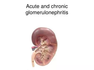
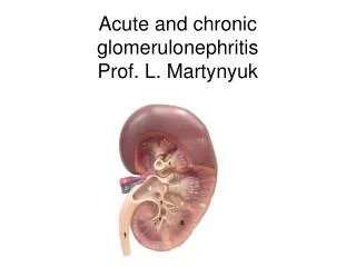
Introduction to nephrology Acute and chronic glomerulonephritis. Internal medicine department №2 Assist. Kvasnitska O.S. The mechanism of urine formation. Acute glomerulonephritis. Etiology. Infectious Streptococcal Nonstreptococcal postinfectious glomerulonephritis Bacterial Viral

E N D
Introduction to nephrologyAcute and chronic glomerulonephritis Internal medicine department №2 Assist. Kvasnitska O.S.
Etiology • Infectious • Streptococcal • Nonstreptococcal postinfectious glomerulonephritis • Bacterial • Viral • Parasitic • Noninfectious • Multisystem systemic diseases • Primary glomerular diseases
Clinical syndromes • urinary (proteinuria ≤1 g/l/24 h, haematuria, leucocyturia), • nephritic (hypertension, gross haematuria, proteinuria 1-3 g/l/24 h, edemas), • nephrotic (proteinuria ≥3,5 g/l/24 h, hypoproteinemia, hypoalbuminemia, hypercholesterolemia, hypercoagulation, edemas), • mixed.
Glomerulonephritis – Diagnostic Tests • CBC (possible anemia, leucocytosis, formula shift to the left, increasing ESR) • Biochemical blood analysis (characterizes the kidney function by the parameters of urea, creatinine, total protein, albumin, serum electrolytes, cholesterol; functional state of the liver (on indicators of ALT, AST, bilirubin) • Examination of the urine (red cells, red-cell casts, nephrotic or sub-nephrotic range proteinuria) • ASO titer (anti streptolysine O) • Kidney scan or biopsy
1= Light yellow, normal colour of urine 2=Light-brown, urine with presence of low proteinuria and microhaematuria 3=Dark-brown, urine with medium presence of proteinuria and microhaematuria 4=Blood-brown, urine with visible haematuria and high level of proteinuria Source: The Internet Journal of Tropical Medicine ISSN: 1540-2681 http://yester.ispub.com/journal/the-internet-journal-of-tropical-medicine/volume-6-number-1/urine-colour-as-a-rapid-assessment-indicator-in-evaluating-the-prevalence-of-schistosoma-haematobium-infection-in-two-endemic-areas-of-benue-state-nigeria.html#sthash.vpY8wWcz.dpuf
Glomerulonephritis –Treatment • Dietary protein is restricted when renal insufficiency (elevated BUN) develop. • Sodium is restricted when the patient has hypertension, edema, and heart failure. • Loop diuretic and antihypertensive medications may be prescribed to control hypertension. • Bed rest during acute phase.
Treatment Treat the underlying infections when acute GN is associated with chronic infections. • Antimicrobial therapy • Antibiotics (eg, penicillin) are used to control local symptoms and to prevent spread of infection to close contacts. • Antimicrobial therapy does not appear to prevent the development of GN, except if given within the first 36 hours. • Loop diuretic therapy • Loop diuretics may be required in patients who are edematous and hypertensive in order to remove excess fluid and to correct hypertension. • Relieves edema and controls volume, thereby helping to control volume-related elevation in BP • Using ACE-ingibitors, AIIRA (angiotensin II receptor antagonists), statins for treatment high BP and lipid abnormalities • Vasodilator drugs (eg, nitroprusside, nifedipine, hydralazine, diazoxide) may be used if severe hypertension or encephalopathy is present
Rapidly progressive glomerulonephritis (RPGN) • Characterized clinically by • A rapid decrease in the GFR of at least 50% over a short period, from a few days to 3 months • The term RPGN was first used to describe a • Group of patients who had an unusually fulminant poststreptococcal glomerulonephritis and a poor clinical outcome • Several years later, • The anti-GBM antibody was discovered to produce a crescentic glomerulonephritis in sheep, and, following this discovery, • The role of anti-GBM antibody in Goodpasture syndrome was elucidated Anti –GBM: antiglomerular basement membrane
RPGN: Pathology • The main pathologic finding is • Extensive glomerular crescent formation • Focal rupture of glomerular capillary walls that can be seen by light microscopy and electron microscopy
Immunological classification: based on the + or - of ANCAs The disorders are also classified based on their clinical presentation RPGN: Classification
RPGN: Classification • Anti-GBM antibody (Approx. 3% of cases) • Goodpasture syndrome (lung and kidney involvement) • Anti-GBM disease (only kidney involvement) • Note: 10-40% of patients may be ANCA positive
RPGN: Classification • Immune complex • Postinfectious (staphylococci/streptococci) • Collagen-vascular disease • Lupus nephritis • Henoch-Schönlein purpura (immunoglobulin A and systemic vasculitis) • Immunoglobulin A nephropathy (no vasculitis) • Mixed cryoglobulinemia • Primary renal disease • Membranoproliferative glomerulonephritis • Fibrillary glomerulonephritis • Idiopathic • Note: Of all patients with crescentic immune complex glomerulonephritis, 25% are ANCA+; < 5% of patients with noncrescentic immune complex glomerulonephritis are ANCA+
RPGN: Classification • Pauci-immune • Wegener granulomatosis (WG) • Microscopic polyangiitis (MPA) • Renal-limited necrotizing crescentic glomerulonephritis (NCGN) • Churg-Strauss syndrome • Note: 80-90% of patients are ANCA+
RPGN: Symptoms *Edema (swelling) of the face, eyes, ankles, feet, legs, or abdomen *Blood in the urine *Dark or smoke-colored urine *Decreased urine volume *Abdominal pain *Cough & Diarrhea *General ill feeling & Fever *Joint aches & Muscle aches *Loss of appetite & Shortness of breath
RPGN: Treatment • Depends on the underlying cause • Corticosteroids may relieve symptoms in some cases • Medications that suppress the immune system may also be prescribed, depending on the cause • Plasmapheresis may relieve the symptoms in some cases • Persons should be closely watched for signs of progression to kidney failure • Dialysis or a kidney transplant may ultimately be necessary
Chronic glomerulonephritis represents the end-stage of all glomerulonephritis with unfavorable evolution. This general (glomerular, vascular and interstitial) affection constitutes the so-called "end stage kidney". In most cases, it is associated with systemic hypertension. (Source: http://www.pathologyatlas.ro/chronic-glomerulonephritis.php)
Minimal-Change Disease • Nil disease or lipoid nephrosis - normal or very mild abnormalities of the glomeruli • Changes can only be seen through electron microscopy. • 90 percent of cases of nephrotic syndrome in children under the age of 10 • More than 50 percent of cases in older children • In adults: use of nonsteroidal antiinflammatory drugs (NSAIDs) • Malignancy: Hodgkin lymphoma Source: http://dc146.4shared.com/doc/tLt3NKse/preview.html Source: http://dc146.4shared.com/doc/tLt3NKse/preview.html
Membranous Nephropathy • Second most common cause of primary nephrotic syndrome in adults • Associated with hepatitis B infection • Autoimmune diseases, thyroid disease, use of certain drugs • Underlying cancer – solid tumor Source: http://dc146.4shared.com/doc/tLt3NKse/preview.html
Focal segmental glomerulosclerosis http://www.nature.com/ki/journal/v68/n4/fig_tab/4496260f1.html
Mesangiocapillary glomerulonephritis • The glomeruli are enlarged, markedly lobulated and ‘rich’ in cells due to proliferation of mesangial cells (more than 2 cells in a plane section) and accumulation of basement membrane-like material. • The capillary walls are thick , their lumens – narrow due to the penetration of mesangium in between the endothelium and basement membrane (so called ‘mesangial interposition’). • With silver stain, this phenomenon is seen as a duplication of basement membrane (‘tram rails’ – metaphor)
Membranoproliferative glomerulonephritis http://www.unckidneycenter.org/kidneyhealthlibrary/mpgn.html
Clinical Manifestations • Uremia-specific findings • Edemas • Hypertension • Jugular venous distension (if severe volume overload is present) • Pulmonary rales (if pulmonary edema is present) • Pericardial friction rub in pericarditis • Tenderness in the epigastric region or blood in the stool (possible indicators for uremic gastritis or enteropathy) • Decreased sensation and asterixis (indicators for advanced uremia)
Characteristics of common glomerular diseases at presentation
Acute Nephritic Syndrome Syndrome characterised in typical cases by: • Haematuria • Proteinuria (1-3 g/l/24 hours) • oliguria • oedema • hypertension • reduced GFR • fluid overload
Nephrotic Syndrome • Proteinuria > 3.5g / 24hrs , due to excessive permeability of glomerular capillary wall. • Leads to hypoproteinemia, hypoalbuminemia, decreased colloid oncotic pressure and edema. • Accompanied by sodium and water retention, hyperlipidemia, vulnerability to infection and thrombotic complications.
Nephrotic syndrome biopsy histology in adults at different ages
Indications to perform renal biopsy • Unexplained renal failure • Acute nephritic syndrome • Nephrotic syndrome • Isolated nonnephroticproteinuria • Isolated glomerularhematuria • Renal masses (primary or secondary) • Renal transplant rejection • Connective-tissue diseases (eg, systemic lupus erythematosus)
Absolute contraindications to renal biopsy include the following: • Uncorrectable bleeding diathesis • Uncontrollable severe hypertension • Active renal or perirenal infection • Skin infection at biopsy site The following are relative contraindications to renal biopsy: • Uncooperative patient • Anatomic abnormalities of the kidney which may increase risk • Small kidneys • Solitary kidney
Treatment of nephrotic syndrome General measures • Dietary sodium restriction • Normal protein intake is advisable. A high-protein diet • (80–90 g protein daily) increases proteinuria and can be harmful in the long term • Infusion of albumin produces only a transient effect. It is only given to diuretic-resistant patients and those with oliguria and uraemia in the absence of severe glomerular damage, e.g. in minimal-change nephropathy. Albumin infusion is combined with diuretic therapy and diuresis often continues with diuretic treatment alone.
Treatment of nephrotic syndrome • The target pressure for patients with proteinuria greater than 1 g/d is less than 125/75 mm Hg; for patients with proteinuria less than 1 g/d, the target pressure is less than 130/80 mm Hg. • Angiotensin-converting enzyme inhibitors (ACEIs) • angiotensin II receptor blockers (ARBs) • combination therapy with ACEIs and ARBs. • Diuretics are often required because of decreased free-water clearance, and high doses may be required to control edema and hypertension when the GFR falls to less than 25 mL/min. • Beta-blockers, calcium channel blockers, central alpha-2 agonists (eg, clonidine), alpha-1 antagonists, and direct vasodilators (eg, minoxidil, nitrates) may be used to achieve the target pressure.
Treatment of nephrotic syndrome • Renal osteodystrophy can be managed early by replacing vitamin D and by administering phosphate binders. • Seek and treat nonuremic causes of anemia, such as iron deficiency, before instituting therapy with erythropoietin. • Discuss options for renal replacement therapy (eg, hemodialysis, peritoneal dialysis, renal transplantation). • Treat hyperlipidemia (if present) • Expose patients to educational programs for early rehabilitation from dialysis or transplantation.
Treatment Minimalchangeglomerulonephritis (MCGN) Acute glomerulonephritis: Prednisolone 1mg/kg/24 hours 4-6 weeks with next reduces of dose and addition of chlorbutin 0,2 mg/kg/24 hours or azathioprine 2 mg/kg/24 hours during of all period of prednisolone receiving In the ineffectiveness of this treatment – cyclosporine A 3-5 mg/kg/24 hours in combination with mild doses of corticosteroids 6-12 weeks Chronic glomerulonephritis: Prednisolone 1mg/kg/24 hours 6-8 weeks to the disappearance or stabilization at the minimum level of proteinuria, further - to 0.5 mg/kg for 4 weeks followed by 5 mg cancellation of in a month. Cytostatics appointed along prednisolone cancellation. Prognosis: 1% progressto ESRF.
Treatment Focal segmental glomerulosclerosis Prednisolone 1mg/kg/24 hours 4-16 weeks. If the full or partial remission achieved, cyclophosphamide added 2 mg/kg or chlorbutin 0.15 mg/kg. In the ineffectiveness of this treatment – prednisolone 0,5 mg/kg every other day and cyclosporine A 2-3 mg/kg during 12 monthes Prognosis: 30–50% progressto ESRF.
Treatment Membranous nephropathy Prednisolone 1mg/kg/24 hours and chlorbutin 0.15 mg/kg or cyclophosphamide 2-3 mg/kg 6-8 weeks In case of relapce or in the ineffectivenes - cyclosporine A 2-3 mg/kg/24 hours 12 monthes in combination with prednisolone 0,5 mg/kg every other day Prognosis: Untreated, 15% complete remission, 9% ESRF at 2–5yrs and 41% at 15yrs
Treatment Mesangiocapillary glomerulonephritis Treatment:None is of proven benefit Prognosis: 50% develop ESRF Mesangial proliferative GN Antibiotics, diuretics, and antihypertensives as necessary. Dialysis is rarely required. Prognosis: Good.
Treatment In the absence of morphological verification of diagnosis Prednisolone 1mg/kg/24 hours 6-8 weeks with subsequent dose reduction and addition of chlorbutin 0.2 mg / kg / day or azathioprine 2 mg / kg / day