Download
1 / 57
830 likes | 2.58k Views
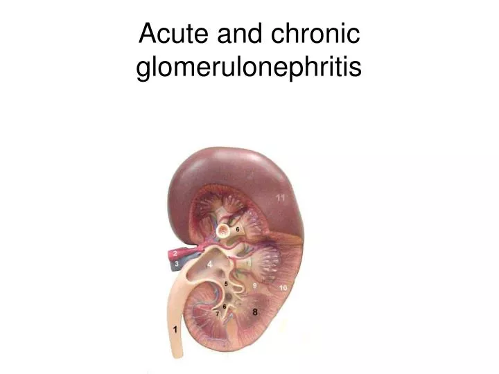
Acute and chronic glomerulonephritis. Glomerulonephritis. Glomerulonephritis – heterogenic group of diseases with primary glomerular localization of pathological changes

E N D
Glomerulonephritis Glomerulonephritis – heterogenic group of diseases with primary glomerular localization of pathological changes It is immunologicaly mediated diffuse inflammatory disease which involves both kidneys symmetrically affecting mainly the glomerulus and associated with changes in tubules, interstitial tissue and vessels.
Etiology of Glomerulonephritis - Beta-hemolytic streptococci of group A ( strains 1, 4, 8, 12, 49). The streptococcal infection is most often in the respiratory tract (pharyngitis, sinusitis, tonsilitis), but infections of other sites (skin and middle car) may also procede nephritis (acute GN) - infections – immunologic factors (infections hepatitis H B3 Ag positive, viruses, rickettsias, mycoplasms) - noninfectious – immunologic factors
Pathogenesis of Glomerulonephritis The two main prosseses are involved in the pathogenesis of glomerulonephritis. 1. Autoimmune: antibodies (antiglomerular basement membrane) react with an antigen in the glomerular basement membrane and produce glomerulonephritis (5% cases). 2. Immune complex theory. Streptococcal or other antigenes provoke antibody response, and the subsequent antigenantibody complexes in the circulation are deposited in the glomerular cappillary walls. These complexes activate the complement pathway with the liberation of chemotactic factors causing polymorpho-leucocytic infiltration the release of lysosomal enzymes from neutrophils and the direct effect of the complement system lead to damage of the capillary wall including the glomerular basal lamina.
Clinical Classification of Gn • ACUTE • Chronic
Clinical Classification of AGn(by L. A. Pyrig 2000) Type urinary syndrome nephrotic syndrome - acute nephritic syndrome Additional information prolonged duration hemature component
Clinical classification of CGn (by L. A. Pyrig, 2000) Type: Urinary syndrome • Nephrotic syndrome Stage: nonhypertensive • Hypertensive • Stage of Chronic renal failure Additional information: • Hematuric component • Phase of activation and remissin
Gistological Classification of Agn(by V. V. Serov, 1983) proliferative glomerulonephritis
Histologic classificationof CGN(by V. V. Serov, 1983) • minimal change disease (lipoid nephrosis • focal and segmental Gn • membranosus Gn • mesangiocapillary Gn (membranoprdiferative Gn) • mesangioproliferative Gn
Membranous nephropathy
Clinical features of AGN (nephritic syndromeTypical clinical picture is presented now rarely. • A latent period of from 5 days to 4-6 weeks occurs between the streptococcal infectious and the abrupt or acute onset of nephritis • Sings of intoxication (fatigue decreased appetite) • Edema (periorbital, leg or sacrlal edema or generalized due to salt and water retention) • Mild or severe hypertension (headaches, visual disturbances secondary to hypertension, rarely hypertensive encephalopathy may be the presenting complains of AGn) • Sings of left ventricular failure (ortopnoe, breathlessness, tachicardia) • Renal impairment manifesting as oliguria or acute renal failure • Dark urine (cola-colored urine) • Changes on the retina (spasm of arteries, dilatation of veins, hemorrhages) • Eclampsia due to cerebral edema and hypertension
Sometimes the onset of the disease may be insidious with weakness, fatigue and malaise or mild edema as the most prominent symptoms after the history of a sore throat, respiratory disease or other • In such situation urinalysis should be prescribed
A history of streptococcal or other infection 1-4 weeks prior to onset of erythrocyturea, proteinurea with development of edema or hypertension are patogonomic of acute glomerulonephritis
Syndromes in GN • Urinary syndrome ( proteinuria (less than 3 gm day), RBCs and casts in the urinary sediment) • Nephritic syndrome (abrupt onset of hematuria, proteinnuria (usually associated with non-nephrogenic range), castiuria, oliguria, hypertension) • Nephrotic syndrome (proteinurea more than 3,5 gm/day, hypoproteinemia and hypoalbuminemia, severe adema, hyperlipidemia) • Edema(mostly is locaried on the face (periorbital), is pale, warm, appears in the morning than decreased, in the second part of the day develops on the legs) • Hypertensive syndrome (hypertension is hyperkinetyc and not severe)
Hypertensive stage: complains on headache, disturbances of vision, insomnia; objective examination reveals high blood pressure, hypertrophy of left ventricular, signs of heart failure, cerebral and cardiac complications • Stage of chronic renal failure signs and symptoms according to the stage (I - IV)
Nephrotic syndrome • - NS –clinical and laboratory syndrome wich includes: • Proteinuriua more than 1 g/m2 24 hours (3,5-4 g/ 24 hours ), • Hypoproteinemia with hypoalbuminemia less than 25 g/l, hyper-alfa-2-globulinemia, • hyperlipiduri, lipiduria, • edema
Complications of NS • nephrotic crises • severe pain in abdomen, associated with peritoneal symptoms, • fever, • oliguria and look like thrombosis of mesenterial arteries and need urgent consultation of surgeon), • skin symptoms migrate erythema
Laboratory findings • Urine analysis • the urine may be scanty, brown, smoky of franky bloody. • From 0.5 to 30 gr/day of protein excreted • The urinary sediment contains RBCs, RBC cast (are the pathognomic of glomerulitis from any etiology) • WBC, renal tubular cells, WBC cast and granular (protein droplets) casts are also may be common • Urinanalysis Nechyporenko (more than 50 000 RBCs in 1 ml of urine is named as hematuric component) • Blood analysis (mild anemia (due to hypervolemia), mild leucocytosis, lympocytosis, increased ESR)
Laboratory findings • Biochemical blood analysis • (hypoproteinemia and hypoalbuminemia, hyperlipidemia (hypoalbuminemia triggers increased synthesis of all forms of plasma proteins including lipoproteins resulting in hyperlipidemia), • elevated level of antistrepolysin-titre (more than 1:3000); • serum complements levels (C3, C4 and the total hemolytic activity) are usually diminished during the active phase of the disease (returns to normal at 6-12 weeks); • serum urea, creatinine may be elevated due to digurea and creatinine clearance reduced; • hypercoagubility may result from • increase urinary loss of antitrombin III • altered levels and/or activity of protein C • hyperfibrunogenemia due to increased hepatic synthesis • impared fibrinolysis • increased platelet aggregability
Instrumental investigation • Renal biopsy usually required for diagnosis in adults • Ultrasound examination may show enlarged kidneys
Parculiaritis of clinical and laboratory signs of CGn according to morphologic changes in kidneys • Mesangioproliferative Gn (Ig A nephropathy) – isolated urinary syndrome, nephritic syndrome, hematuria in adults • Mesangiocapillary Gn – nephrotic syndrome, urinary syndrome with hematuric component, hypertension • Membranosus Gn – nephrotic syndrome (80%) in age 40-50 • Focal and segmental Gn - nephrotic syndrome, hypertension in Afro-Americans • Minimal change disease - nephrotic syndrome in children • Fibroplastic Gn - nephrotic syndrome (50%), hypertension, chronic renal failure
Differential diagnosis Urinary syndrome • Acute pyelonepritis • Activation of primary chronic glomerulonephritis • Toxic nephritis • Goodpasture’s Syndrome • Hereditary nephritis (Alport’s Syndrome) Nephrotic syndrome • Amyloidosis • Diabetic nephropathy • Colagenic nephtopathy (SLE scleroderma) Hematuric component • Malignancy associated nephritis • Urotuberculosis • Renal stones
An example of diagnosis • Acute glomerulonephritis, urinary syndrome, hematuric component • Acute glomerulonephritis, nephrotic syndrome
An example of diagnosis • Chronical glomerulonephritis, urinary syndrome, hypertensive stage, phase of activation
Duration of acute glomerulonephritis • Recovering during first 2-4 weeks or 2-3month • Prolonged duration (duration more than 4 month, full recovering is 2-3 times rare) • Negative prognostic feature is nephrotic syndrome, associated with severe hypertension • Development of chronic glomerulonphritis (urinary syndrome, edema or hypertension are present more than 12 month)
Complications of acute glomerulnepherts • Eclampsia (angiospastic encephalopathy) • Acute heart (left ventricular) failure • Acute renal failure
Treatment of acute glomerulonephritisAcute glomerulonephritis have to be treated only in speciallised nephrologic department • Regimen: bed-rest during 2-4-6 weeks until desappearing of edema and normalizing of blood pressure • Diet № 7a • Daily record of fluide intake and output • Restriction of dietary protein if azotemia and metabolic acidosis are present • Salt free diet (Sodium intake is restricted only when circulation overload, edema, or severe hypertension is present)
The aimof drug therapyis recovering of the patient • Anti microbal drug • Symptomatic therapy • Membranenostabilizative therapy • Pathogenetic therapy
Antimicrobal therapy • If a bacterial infection is still present when nephritis is discovered, it should be treated with an appropriated antimicrobial drug • Semisynthetic penicillins in middle therapeutic doses have to be prescripted
Symptomatic therapyEdema • Loop diuretics such as furosemide or lasix (40-400 mg/day or 1-2 gr/day) help in the management of the expanded extracellular fuid volume (side effects: hypocholremic alkalosis, decreasing K, Na level inblood) • In patients with decreased of furosemide should be prescribed uregit (50-200-500 mg/day orally) or the combination with the thiazides (hypothiazide 25-100 mgm/day) • 2.4 % solution euphylline 10 ml i/v • Albumin may help in the management of hypoproteinemia • Daily weighting to check change in the body fluid status and record of fluid intake and output have to be made in patients which receive diuretics
Hypertensive syndrome Antihypertensive drug therapy is usually started with single drug, but if there is incomplete response a second drug is added. One of the following drugs as a single drug treatment can be used: • ACE inhibitors(or angiotensine II reseptors blockers) • (loop) diuretics • calcium channel blockers (non dihydropiridine agents) If single drug treatment is unsuccessful then the combination therapy may be given as two-drugs or three – drugs therapy Two – drug therapy: • calcium channel blocker + diuretic • ACE inhibitor + diuretic Triple – drug therapy is used very rare • calcium channel blocker + diuretic + ACE inhibitor Such drugs as adelfan or trirezide (which contained fixed doses of several hypotensive drugs) are not good in therapy of hypertensive syndrome
Hematuric component • Dicinon (etamsilate) 2 ml 12.5% solution twice a day (7-10 days) i/m, then 0.25-0.5 three times a day orally • Kvarcetin 1.0 in a half of glass of water three times a day. • Ascorbinic acide 500 mg a day. • Ascorutine 1 tabl. three times a day • Rutine and other.
Membranostabilizative therapy • Have to be prescribed in patients with AGn, urinary syndrome, hematuric component, after prescription of symptomatic therapy. • Unitiol (5 ml 5% solution i/m during 1 month) • Dimephosphon (100 mkg/kg/day 1 month) • Aminochinolytic drugs (delagil – 0.25 two times a day orally 1 month, then 0.25 a day during 5-12 month) • (side effects: leucopenia, degeneration of retina, allergy, dyspepsia) • ά-tocoferol ( 50 mgm/day – 5-12 month)
Pathogenetic therapyhave to be used in patients with: • Gn, nephrotic syndrome after 3-4 weeks from the beginning of the disease, when symptomatic and membranostabilisative therapy is unsuccessful
Pathogenetic therapy includes: • Glucocorticoids • Cytostatics • Anticoagulants and antiagregative drugs
Glucocorticosteroids • Prednisolone 1 mg/kg/day for 4-6 weeks followed by decreasing of dosage on 2.5 mg each 5-7 days • In patients with high activity of patogenetic process pulse-therapy with metylprednisolone (metipred, soly-pred, solu-medrol) (1000 mg/d three days) can be used and then therapy in previous doses • (Side effects: obesity, hirsutism, disturbances of menstrual function, achne, Cushing syndrome, ulcers of alimentary tract, hyperglycemia, hemorrhagic, pancreatitis, psychiatric disturbances. • After abrupt discontinuoing of the drug usage can be worsening of the duration of the main disease)