Download
1 / 29
290 likes | 329 Views
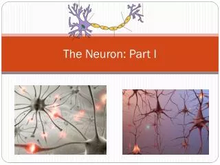
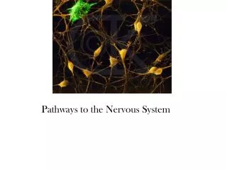
The Neuron. Units of Function and the Nervous System. What is the neuron?. The neuron , or nerve cell, sends and receives signals that affect many aspects of behavior and motor control. Three Major Structures of the Neuron.

E N D
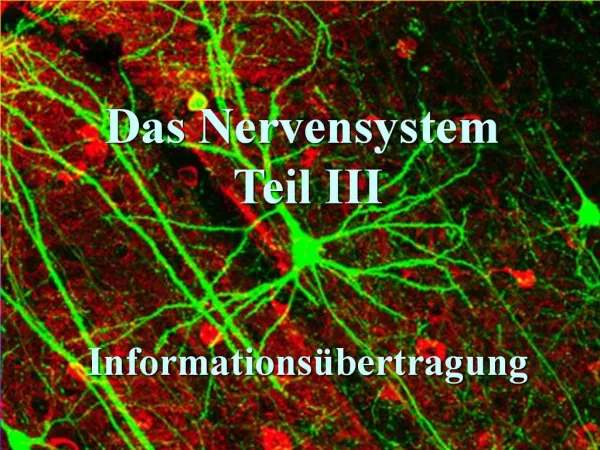
The Neuron Units of Function and the Nervous System
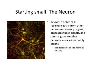
What is the neuron? The neuron, or nerve cell, sends and receives signals that affect many aspects of behavior and motor control.
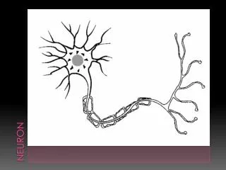
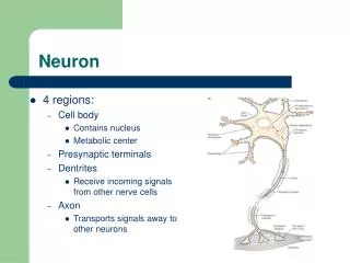
Three Major Structures of the Neuron The cell body (or soma) contains cytoplasm and the nucleus, which includes the chromosomes. Mitochondria (the powerhouse of the cell) in the cell body perform metabolism. Ribosomes synthesize proteins. Extending outward from the soma are dendrites (Greek=little trees), the receiving/input branches of the neuron. The axon emerges from the soma as a single conducting fiber.
More about the Axon The axon branches and ends in tips called presynaptic terminals (also known as terminal buttons, boutons, or telodendria). Neurotransmitters are stored in structures of the presynaptic terminal known as vesicles. The axon may be covered by an insulating myelin sheath (think insulation on an electrical cord), made of specialized cells such as oligodendrocytes (central nervous system, or CNS) and Schwann cells categorized as glial cells (peripheral nervous system, or PNS). Glial cells (from Greek word meaning glue”) were once thought of as cells that simply hold the neurons together. We know now that they play important roles in neural function. Spaces between segments of myelin are called nodes of Ranvier. One function of the myelin sheath is to speed up neural impulses.
The Neurotransmitters Neurotransmitters are chemical “messengers” used to pass a variety of messages between the neurons. Make sure you know what each type does!
Acetylcholine Acetylcholine (ACh) causes contraction of the skeletal muscles, helps regulate heart muscles, promotes arousal in the brain, and transmits messages between the brain and spinal cord. Low ACh means low arousal and low attention; its depletion is associated with Alzheimer’s disease.
Glutamate Glutamate and aspartate stimulate receptors associated with learning and memory, as well as many sensory and content outline 8 motor functions. Glutamate is the most abundant excitatory neurotransmitter in the brain. An overabundance of glutamate may lead to migraine headaches; often connected with MSG (monosodium glutamate) in foods, a result of overstimulation.
Gamma-Aminobutyric Acid GABA (gamma-aminobutyric acid) inhibits the firing of neurons. It is the most abundant inhibitory neurotransmitter in the brain. GABA is associated with calming effects. A lack of GABA is connected to seizures, tremors, insomnia, anxiety, epilepsy, and Huntington’s disease.
Dopamine Dopamine (DA) is primarily involved in processing smooth and coordinated gross motor movements and in attention, learning, and reinforcing effects of several often-abused drugs. Parkinson’s disease is associated with the death of DA-producing neurons. Dopamine release in the nucleus accumbens is linked to addictive drugs, sex, and attention grabbing experiences in general, including video game playing.
Norepinephrine Norepinephrine (NE) is found in neurons in the autonomic nervous system (ANS). NE governs sympathetic arousal by activating the heart and blood vessels, thus giving rise to the “fight or flight” syndrome as well as other excitatory actions. It is also released in the brain to enhance attention and memory for emotionally charged events.
Serotonin Serotonin (SE or 5-HT, for 5-hydroxytryptamine) plays a role in the regulation of mood; control of eating, sleep, arousal; the regulation of pain; and control of dreaming. Most of the drugs that relieve depression increase activity at serotonergic synapses.
Opioid Peptides Opioid peptides, such as endorphins, are often considered the brain’s own painkillers. These are endogenous chemicals that modulate the experience of pain or pleasure.
Peptide Neuromodulators Dozens of peptide neuromodulators are released by small groups of brain neurons. These peptides produce long-lasting, widespread effects, much like hormones, to alter hunger, thirst, and other long-lasting behaviors.
The Neural Impulse The Neural impulse, known as the action potential, is electrochemical (uses both electrical and chemical systems to pass information). It is an all-or-nothing action, as an axon either fires or does not. https://www.youtube.com/watch?v=4M1zzT9J_y4
The Action Potential A resting neuron is more negative inside the cell membrane than outside. The resting neural membrane potential is about -70mV (on average). (Physiologists refer to voltage differences as “potential” because they represent potential energy.) When sufficiently stimulated to threshold, the cell membranes admit sodium, creating a depolarization (loss of potential) that “kicks” the electrical charge down the axon to the presynaptic terminals. The brief change in potential is called the action potential.
The Action Potential & the Sodium-Potassium Pump The action potential reverses the charge across the membrane—it changes from -70 mV to about +50 mV for about 1 msec. After the peak of the action potential, the sodium channels close. However, the potassium channels are open wider than usual, and potassium exits from the cell to the outside, carrying a positive charge and returning the membrane to its original condition (or a slightly increased hyperpolarization). This transfer of sodium and potassium ions down the length of the neuron is referred to as the sodium-potassium pump.
Action Potential Speed The more intense a stimulus, the more frequently a neuron fires, but the amplitude and velocity of the action potentials do not change. (Your textbook compares this to the firing of a bullet: you can pull the trigger of a gun faster to fire more bullets, but you can’t pull the trigger harder to make the bullet fly faster.) When the axon is myelinated (has a myelin sheath), conduction speed is increased. Depolarizations occur only at the nodes of Ranvier, and, therefore, the action potential jumps from one node to the next. This is called saltatory conduction. (So, impulses travel faster through a myelin sheathed neuron).
End of the Line The message now arrives at the presynaptic terminals and prepares to release neurotransmitters (chemicals) from sacs (vesicles). After a brief period of time—the refractory period—the neuron is ready to fire again. Let’s have Hank Green take us through a summary: https://www.youtube.com/watch?v=OZG8M_ldA1M
The Synapse Neurons signal by transmitting chemical messages to adjacent neurons, gland cells, or muscle cells (synaptic transmission). Tiny gaps between neurons are called synaptic clefts. A particular terminal button of an axon, the synaptic cleft itself, and the receiving portion of another neuron, gland cell, or muscle cell together constitute the synapse.
The Synapse When action potentials arrive at the terminal, they trigger the release of neurotransmitter molecules from synaptic vesicles into the synaptic cleft. A signal is transmitted from one neuron to the next when the neurotransmitter molecules from the presynaptic neuron bind with the postsynaptic neuron (at its dendrites), much like a “lock-and-key” mechanism, thus changing the potential of the postsynaptic neuron.
To fire or not to fire If the binding of the neurotransmitter to the postsynaptic receptor site makes the neuron more likely to fire, the effect is called excitatory. If the binding of the neurotransmitter to the postsynaptic receptor site prevents or lessens the likelihood of the firing of the neuron, the effect is called inhibitory. More Hank! https://www.youtube.com/watch?v=VitFvNvRIIY
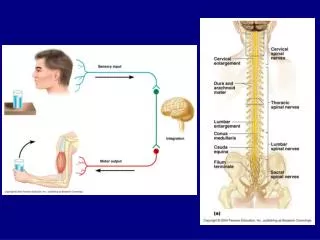
Reflexes The simplest form of behavior, called a reflex, involves impulse conduction over a few neurons. This path is called a reflex arc. Sensory or afferent neurons transmit impulses from sensory receptors to the spinal cord or brain. Motor or efferent neurons conduct impulses (motor commands) away from the brain or spinal cord to the muscles and glands. Muscle and gland cells acting on motor commands are called effectors. Interneurons, located entirely within the brain and spinal cord, intervene between one neuron and another. It relays signals between afferent and efferent neurons.