Download
1 / 9
90 likes | 242 Views
THE NEURON. PREPARED BY HUGH POTTER,Ph.D BIOLOGY DEPARTMENT UNION COUNTY COLLEGE. ALL IMAGES USED WITH THE PERMISSION OF THE PUBLISHERS OF THE SLICE OF LIFE VIDEO DISK. THE NEURON. d. cb. a. d. d.

E N D
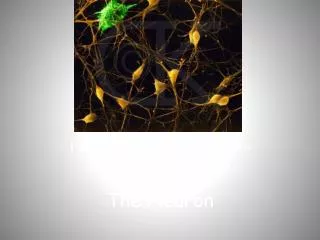
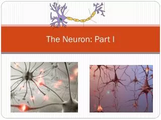
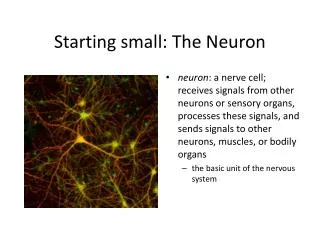
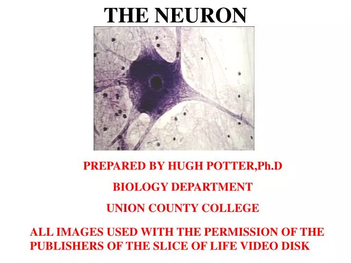
THE NEURON PREPARED BY HUGH POTTER,Ph.D BIOLOGY DEPARTMENT UNION COUNTY COLLEGE ALL IMAGES USED WITH THE PERMISSION OF THE PUBLISHERS OF THE SLICE OF LIFE VIDEO DISK
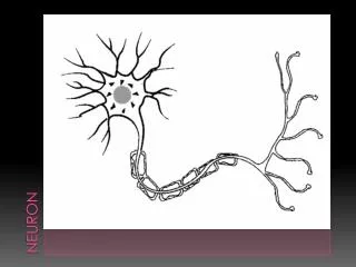
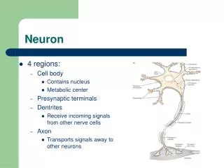
THE NEURON d cb a d d The cell body (perikaryon) is the portion of the nerve cell that surrounds the nucleus. The size of a typical neuron cell body varies from 4 to 135um. Multipolar neurons have several branches arising form the cell body (cb). There is usually one axon (a) and many dendrites (d). Dendrites carry nerve impulses to the cell body. The axon is designed to carry nerve messages away from the cell body.
NISSLE BODIES Nissle bodies(arrows) are widely distributed in the cytoplasm of most neurons. They are more numerous in motor neurons. Nissle bodies are formed by clumps of ribosomes attached to portions of endoplasmic reticulum. They signify a high level of protein synthesis
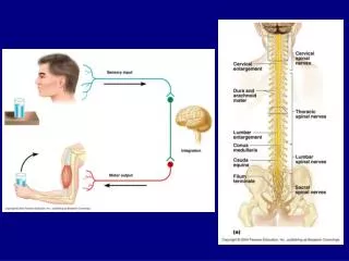
UNIPOLAR NEURONSIn A DORSAL ROOT GANGLION * * * * * Unipolar cells have a globular cell body (asterisks) with a single branch that divides. One of these divisions carries nerve impulses into the cell body from receptor tissue. The other division passes these impulses onto the spinal cord. Cell bodies of this type of neuron can be found in the dorsal root ganglia near the spinal cord.
MYELINATED AXON All nerve cell processes in the central nervous system (CNS) peripheral nervous system (PNS) have one or more sheaths or coverings. Myelinated processes in the PNS have a sheath of Schwann cells around a myelin wrapping. The myelin is produced by the Schwann cells.
NODES OF RANVIER The myelin sheath is seen to be interrupted every 0.1 micron to 1.5 mm by a gap called the Node of Ranvier (arrows). The jumping of the nerve message from node to node speeds up the passage of the nerve impulse along myelinated axons.
NEUROFILAMENTS Neurofilaments (arrows) are delicate fibers present throughout the cytoplasm of the cell body and extending in bundles into dendrites and axons. These bundles can extend from the cell body to the ends of cell processes. Neurofibrils are composed of microtubules formed from the protein tubulin and microfilaments of actin and myosin. They are responsible for supporting the internal framework of the cell, as well as, transport of substances within the cell.
END BRANCHES AND MOTOR END PLATES The axon (a) is seen to divide into many telodendria (t) (end branches). At the terminus of each end branch is a synaptic knob (arrow). The motor end plates are under the knob on the surface of the skeletal muscle fiber. a t
The synapse is located at the end of each axonal end branch. Here the end branch forms a small synaptic knob (sk). This knob is adjacent to a tiny cleft or synapse (s). When a nerve impulse reaches this knob, a drug called a neurotransmitter is released from vesicles into the synapse The neurotransmitter diffuses across the gap and binds toreceptors on the membrane of the adjacent neuron or muscle cell. This will initiate an electrical change in the membrane of the adlacent neuron leading to a nerve impulse in that cell. SYNAPTIC KNOB sn s s