Download
1 / 1
30 likes | 189 Views
Fiber Optic Sample Arm for Raman Spectroscopy (RS) and Optical Coherence Tomography (OCT) Probe John Acevedo, Chris Miller, and Kelly Thomas. 8”. 5 ”. Distance to Image vs. Beam Diameter. Gaussian Beam diameter( μ m). Distance to Image ( μ m). Motivation. Methods. Results.

E N D
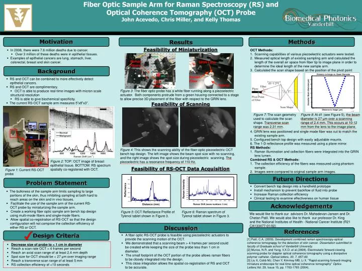
Fiber Optic Sample Arm for Raman Spectroscopy (RS) and Optical Coherence Tomography (OCT) Probe John Acevedo, Chris Miller, and Kelly Thomas 8” 5” Distance to Image vs. Beam Diameter Gaussian Beam diameter(μm) Distance to Image (μm) Motivation Methods Results Feasibility of Miniaturization • In 2008, there were 7.6 million deaths due to cancer. • Over 3 million of these deaths were in epithelial tissues. • Examples of epithelial cancers are lung, stomach, liver, • colorectal, breast and skin cancer. OCT Methods: Scanning capabilities of various piezoelectric actuators were tested. Measured optical length of existing sampling arm and calculated the length of the overall air space from fiber tip to image plane in order to determine the ideal length of the new sample arm. Calculated the scan shape based on the position of the pivot point 4. GRIN lens was positioned and single mode fiber was cut to match the existing sample arm. 5. Configured bench top design with easily adjustable mounts. 6. The 1-D reflectance profile was measured using a plane mirror. RS Methods: Raman illumination and collection fibers were integrated into the GRIN lens lumen. Combined RS & OCT Methods: The collection efficiency of the fibers was measured using phantom sample. Images were compared to original sample arm images. Raman Fibers GRIN Lens Background Raman Fiber OCT Fiber • RS and OCT can be combined to more effectively detect • epithelial cancers. • RS and OCT are complimentary. • OCT is able to produce real-time images with micron scale structural resolution • RS is able to give biochemical specificity. • The current RS-OCT sample arm measures 5”x8”x3”. Tylenol Figure 3: The fiber optic probe has a white fiber running along a piezoelectric actuator. Both components protrude from a green housing connected to a stage to allow precise 3D placement of the fiber with respect to the GRIN lens. Feasibility of Scanning Figure 7: The scan geometry used to calculate the scan shape. Transverse scan range was 2.31 mm. • Figure 8: At d1 (see Figure 5), the beam diameter is 27 μm over a scanning range of 2.4 mm. This occurs at 10-12 mm from the lens to the image plane. Figure 4: This shows the scanning ability of the fiber-optic piezoelectric OCT bench top design. The left image shows the beam spot size with no scanning, and the right image shows the spot size during piezoelectric scanning. The piezoelectric has a resonance frequency of 110 Hz. Figure 2: TOP: OCT image of breast epithelial tissue. BOTTOM: RS spectrum spatially co-registered with OCT. Feasibility of RS-OCT Data Acquisition Figure 1: Current RS-OCT probe Problem Statement FutureDirections • Convert bench top design into a handheld prototype • Install mechanism to prevent backflow of fluid into probe • Increase Raman collection efficiency • Clinical testing to examine effectiveness on human tissue • The bulkiness of the sample arm limits sampling to large • portions of the skin, thus inhibiting sampling at both hard to • reach areas on the skin and in vivo tissues. • Facilitate the use of the sample arm of the current RS- • OCT probe by miniaturizing the sample arm. • Create a working fiber optic sample arm bench top design • using multi-mode fibers and single-mode fibers. • Allow spatial co-registration of RS-OCT so that the design • configuration will not comprise the collection efficiency of • either RS or OCT. Acknowledgements Figure 5: OCT Reflectance Profile of Tylenol tablet shown in Figure 3. Figure 6: Raman spectrum of Tylenol tablet shown in Figure 3. We would like to thank our advisors Dr. Mahadevan-Jansen and Dr. ChetanPatil. We would also like to thank our professor Dr. King and the National Institutes of Health - National Cancer Institute (R21 CA133477-01/02) Design Criteria Discussion References • A fiber optic RS-OCT probe is feasible using piezoelectric actuators to • provide the scanning motion of the OCT. • We demonstrated that a scanning beam > 4 frames per second could • be created while keeping the size of the probe less than 1 cm in • diameter. • The small footprint of the OCT portion of the probe allows raman fibers • to be closely integrated into the design. • This close integration allows the spatial co-registration of RS and OCT • to be accurate. [1] Patil, C.A. (2009). Development combined raman spectroscopy-optical coherence tomograpgy for the detection of skin cancer. Dissertation submitted to faculty of Graduate school of Vanderbilt University. [2] Munce, N.R. and Yang, V.X.D. et al.(2008). Electrostatic forward-viewing scanning probe for doppler optical coherence tomography using a dissipative polymer catheter. Optical letters, 33, 7, 657-60. [3] Liu X, Cobb MJ, Chen Y, Kimmey MB, Li X. "Rapid-scanning forward-imaging miniature endoscope for real-time optical coherence tomography" Optics Letters,Vol. 29, Issue 15, pp. 1763-1765 (2004). • Decrease size of probe to < 1 cm in diameter • Reach a scan rate OCT > 4 frames per second • Reach an axial scan range of at least 2.4 mm depth • Spot size for OCT should be < 27 μm over imaging range • Reach a transverse scan range of at least 5 mm • RS collection efficiency of <10 seconds