Download
1 / 24
450 likes | 1.62k Views
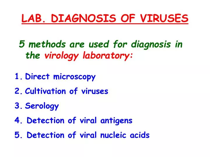
LAB. DIAGNOSIS OF VIRUSES 5 methods are used for diagnosis in the virology laboratory: Direct microscopy Cultivation of viruses Serology 4 . Detection of viral antigens 5. Detection of viral nucleic acids. DIRECT MICROSCOPY @ Three procedures can be used :

E N D
LAB. DIAGNOSIS OF VIRUSES • 5 methods are used for diagnosis in the virology laboratory: • Direct microscopy • Cultivation of viruses • Serology • 4. Detection of viral antigens • 5. Detection of viral nucleic acids
DIRECT MICROSCOPY @ Three procedures can be used : 1. Light microscopy: to reveal inclusion bodies or multinucleated giant cells e.g. herpesvirus 2. Immunofluorescence microscopy: to readviral fluorescent antibodies 3. Electron microscopy: to detect virus particles
Cultivation of Viruses • Inoculation of fertile: Specimen is injected in 10-12 day-old eggs. Three days later eggs are opened to see spots on membranes and fluids. • Inoculation of lab. Animals: Specimen is injected in 2 day-old suckling mice, and look for clinical signs in mice. • 3. Viral tissue culture
VIRAL TISSUE CULTURE @ Transport specimen in Hank’s medium or store at 4 °C immediately @ Virus growth in living cells produces a Cytopathic Effect (CPE) . @ Identification depends on : * time taken for the CPE to appear * type of cell present in CPE
If no CPE, virus is detected by: • Hemadsorption Test: For viruses with hemagglutination antigens, eg mumps, and influenza viruses • 2. Interference Test:Formation of CPE by another virus, eg Rubella which does not cause CPE, but it can cause CPE when ECHO virus is added to the cell culture
3. Phenol red indicator changes yellow:This is due to decrease in acid production when the infectedcells die in medium 4. Phenol red indicator remains red (alkaline): This is used to detect Eenteroviruses
5. Use of a known antibody and made to react with the virus antigen in the cell culture by one of the following tests : • 1) Complement Fixation Test . • 2) Hemagglutination Inhibition Test • 3) Neutralization Test. • 4) Fluorescent-Antibody Test
5) Radioimmunoassay Test. 6) Enzyme-Linked Immunosorbent Assay (ELlSA) Test. 7) Immunoelectron microscopy Test
SEROLOGY @ Blood is collected twice: at the acute stage and 10-14 days later @ If the antibody titer in second sample is at least 4-fold higher than the titer in the acute stage sample, patient is considered to be infected.
@ IgM antibody only is used to diagnose • acute infection, e g infection by • hepatitis B virus. • @ Tests used are: Complement fixation, Hemagglutination-inhibition, ELlSA, Neutralization, Radioimmunoassay, and • Fluorescent-antibody tests • @ Heterophile non-specific agglutination • antibody test: can be used to detect • Epstein-Bar virus
DETECTION OF VIRAL ANTIGENS @ Detected in patient's blood or body fluids by ELISA. @ Examples : * p24 antigen of HIV * HBs antigen of Hepatitis B
DETECTION OF VIRAL NUCLEIC ACIDS @ DNA,RNA, & mRNA are detected in blood or tissues by polymerase chain reaction (PCR), using DNA or RNA probe @ For small amounts of N.A., amplify these N.A. by PCR.