Download

1 / 72
750 likes | 921 Views
5 The Integumentary System. An Introduction to the Integumentary System. Learning Outcomes 5-1 Describe the main structural features of the epidermis, and explain the functional significance of each. An Introduction to the Integumentary System. Learning Outcomes

E N D
5 The Integumentary System
An Introduction to the Integumentary System • Learning Outcomes • 5-1 Describe the main structural features of the epidermis, and explain the functional significance of each.
An Introduction to the Integumentary System • Learning Outcomes • 5-5 Describe the structure and functions of the dermis. • 5-6 Describe the structure and functions of the hypodermis. • 5-7 Explain the structural basis for hair texture and color.
An Introduction to the Integumentary System • Learning Outcomes • 5-8 Discuss the various kinds of glands in the skin, and list the secretions of those glands. • 5-9 Describe the anatomical structure of nails, and explain how they are formed.
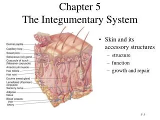
An Introduction to the Integumentary System • The Integument • Is the largest system of the body • 16% of body weight • 1.5 to 2 m2 in area • The integument is made up of two parts • Cutaneous membrane (skin) • Accessory structures
An Introduction to the Integumentary System • Two Components of the Cutaneous Membrane • Outer epidermis • Superficial epithelium (epithelial tissues) • Inner dermis • Connective tissues
An Introduction to the Integumentary System • Accessory Structures • Originate in the dermis • Extend through the epidermis to skin surface • Hair • Nails • Multicellular exocrine glands
An Introduction to the Integumentary System • Connections • Cardiovascular system • Blood vessels in the dermis • Nervous system • Sensory receptors for pain, touch, and temperature
An Introduction to the Integumentary System • Hypodermis (Superficial Fascia or Subcutaneous Layer) • Loose connective tissue • Below the dermis • Location of hypodermic injections
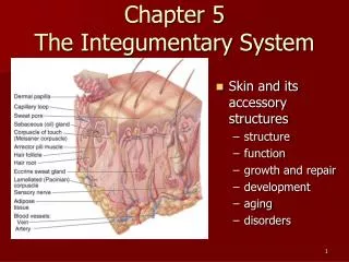
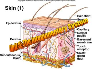
Figure 5-1 The Components of the Integumentary System Accessory Structures Cutaneous Membrane Hair shaft Epidermis Pore of sweatgland duct Papillary layer Tactile corpuscle Dermis Reticular layer Sebaceous gland Arrector pili muscle Sweat gland duct Hair follicle Lamellated corpuscle Hypodermis Nerve fibers Sweat gland Artery Cutaneousplexus Vein Fat
Figure 5-1 The Components of the Integumentary System Cutaneous Membrane Epidermis Papillary layer Dermis Reticular layer Hypodermis
Figure 5-1 The Components of the Integumentary System Accessory Structures Hair shaft Pore of sweatgland duct Tactile corpuscle Sebaceous gland Arrector pili muscle Sweat gland duct Hair follicle Lamellated corpuscle Nerve fibers Sweat gland Artery Cutaneousplexus Vein Fat
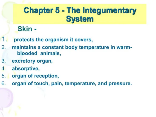
An Introduction to the Integumentary System • Functions of Skin • Protectionofunderlying tissues and organs • Excretion of salts, water, and organic wastes (glands) • Maintenance of body temperature (insulation and evaporation) • Productionof melanin
An Introduction to the Integumentary System • Functions of Skin • Productionof keratin • Synthesisof vitamin D3 • Storage of lipids • Detection oftouch, pressure, pain, and temperature
5-1 Epidermis • The Epidermis • Is avascular stratified squamous epithelium • Nutrients and oxygen diffuse from capillaries in the dermis
Figure 5-2 The Basic Organization of the Epidermis Stratumcorneum Epidermis Stratumlucidum Epidermalridge Basement membrane Dermalpapilla Dermalpapilla Dermis Epidermalridge Dermis Thin skin LM 154 Thick skin LM 154
Figure 5-2a The Basic Organization of the Epidermis Epidermis Epidermalridge Dermalpapilla Dermis The structural relationship andinterface between the epidermisand underlying dermis. Theproportions of the various layersdiffer with the location sampled.
5-1 Epidermis • Cells of the Epidermis • Keratinocytes • Contain large amounts of keratin • Are the most abundant cells in the epidermis
5-1 Epidermis • Thin Skin • Covers most of the body • Has four layers of keratinocytes • Thick Skin • Covers the palms of the hands and soles of the feet • Has five layers of keratinocytes
Figure 5-2b The Basic Organization of the Epidermis Stratumcorneum Basement membrane Dermis LM 154 Thin skin A micrograph ofthin skin, which covers most ofthe exposedbody surface.
Figure 5-2c The Basic Organization of the Epidermis Stratumcorneum Stratumlucidum Dermalpapilla Epidermalridge Thick skin LM 154 A micrograph of thickskin, which coversthe surface of thepalms and the solesof the feet.
5-1 Epidermis • Structures of the Epidermis • The five strata of keratinocytes in thick skin • From basal lamina to free surface • Stratum basale • Stratum spinosum • Stratum granulosum • Stratum lucidum • Stratum corneum
Figure 5-3 The Structure of the Epidermis Surface Stratumcorneum Stratumlucidum Stratumgranulosum Stratumspinosum Stratum basale Basementmembrane Papillary layer of dermis Dermis Thick skin LM 210
5-1 Epidermis • Stratum Basale • Is attached to basement membrane by hemidesmosomes • Forms a strong bond between epidermis and dermis • Forms epidermal ridges (e.g., fingerprints) • Dermal papillae (tiny mounds) • Increase the area of basement membrane • Strengthen attachment between epidermis and dermis • Has many basal cells or germinative cells
Figure 5-4 The Epidermal Ridges of Thick Skin Pores of sweatgland ducts Epidermalridge Thick skin SEM 25
5-1 Epidermis • Specialized Cells of Stratum Basale • Merkel cells • Found in hairless skin • Respond to touch (trigger nervous system) • Melanocytes • Contain the pigment melanin • Scattered throughout stratum basale
5-1 Epidermis • Stratum Spinosum — the“spiny layer” • Produced by division of stratum basale • Eight to ten layers of keratinocytes bound by desmosomes • Cells shrink until cytoskeletons stick out (spiny) • Continue to divide, increasing thickness of epithelium • Contain dendritic (Langerhans) cells, active in immune response
5-1 Epidermis • Stratum Granulosum — the “grainy layer” • Stops dividing, starts producing • Keratin • A tough, fibrous protein • Makes up hair and nails • Keratohyalin • Dense granules • Cross-link keratin fibers
5-1 Epidermis • Cells of Stratum Granulosum • Produce protein fibers • Dehydrate and die • Create tightly interlocked layer of keratin surrounded by keratohyalin
5-1 Epidermis • Stratum Lucidum —the “clear layer” • Found only in thick skin • Covers stratum granulosum
5-1 Epidermis • Stratum Corneum —the “horn layer” • Exposed surface of skin • 15 to 30 layers of keratinized cells • Water resistant • Shed and replaced every 2 weeks
5-1 Epidermis • Keratinization • The formation of a layer of dead, protective cells filled with keratin • Occurs on all exposed skin surfaces except eyes • Skin life cycle • It takes 15–30 days for a cell to move from stratum basale to stratum corneum
5-1 Epidermis • Perspiration • Insensible perspiration • Interstitial fluid lost by evaporation through the stratum corneum • Sensible perspiration • Water excreted by sweat glands • Dehydration results: • From damage to stratum corneum (e.g., burns and blisters [insensible perspiration]) • From immersion in hypertonic solution (e.g., seawater [osmosis])
5-1 Epidermis • Hydration • Results from immersion in hypotonic solution (e.g., freshwater [osmosis]) • Causes swelling of epithelial cells, evident on the palms and soles
5-5 The Dermis • The Dermis • Located between epidermis and subcutaneous layer • Anchors epidermal accessory structures (hair follicles, sweat glands) • Two components • Outer papillary layer • Deep reticular layer
5-5 The Dermis • The Papillary Layer • Consists of areolar tissue • Contains smaller capillaries, lymphatics, and sensory neurons • Has dermal papillae projecting between epidermal ridges
5-5 The Dermis • The Reticular Layer • Consists of dense irregular connective tissue • Contains larger blood vessels, lymphatic vessels, and nerve fibers • Contains collagen and elastic fibers • Contains connective tissue proper
5-5 The Dermis • Dermatitis • An inflammation of the papillary layer • Caused by infection, radiation, mechanical irritation, or chemicals (e.g., poison ivy) • Characterized by itch or pain
5-5 The Dermis • Innervation of the Skin • Nerve fibers in skin control: • Blood flow • Gland secretions • Sensory receptors • Light touch—tactile corpuscles, located in dermal papillae • Deep pressure and vibration—lamellated corpuscles, in the reticular layer
5-6 The Hypodermis • The Hypodermis (Subcutaneous Layer) • Lies below the integument • Stabilizes the skin • Allows separate movement • Made of elastic areolar and adipose tissues • Connected to the reticular layer of integument by connective tissue fibers • Few capillaries and no vital organs • The site of subcutaneous injections using hypodermic needles
5-6 The Hypodermis • Deposits of Subcutaneous Fat • Distribution patterns determined by hormones • Reduced by cosmetic liposuction (lipoplasty)
5-7 Hair • Hair, Hair Follicles, Sebaceous Glands, Sweat Glands, and Nails • Integumentary accessory structures • Derived from embryonic epidermis • Located in dermis • Project through the skin surface
5-7 Hair • Human Body • The human body is covered with hair, except: • Palms • Soles • Lips • Portions of external genitalia
5-7 Hair • Functions of Hair • Protects and insulates • Guards openings against particles and insects • Is sensitive to very light touch
5-7 Hair • The Hair Follicle • Located deep in dermis • Produces nonliving hairs • Wrapped in a dense connective tissue sheath • Base is surrounded by sensory nerves (root hair plexus)
5-7 Hair • Accessory Structures of Hair • Arrector pili • Involuntary smooth muscle • Causes hairs to stand up • Produces “goose bumps” • Sebaceous glands • Lubricate the hair • Control bacteria
5-7 Hair • Regions of the Hair • Hair root • Lower part of the hair • Attached to the integument • Hair shaft • Upper part of the hair • Not attached to the integument
Figure 5-10a Hair Follicles and Hairs Exposedshaftof hair Sebaceousgland Arrectorpilimuscle Connectivetissue sheath Root hairplexus Single hair follicle, showing the associated accessorystructures; a superficialview of the deeper portionsof the follicle illustrates theconnective tissue sheathand the root hair plexus.
Figure 5-10b Hair Follicles and Hairs Hair Structure The cortex contains thicklayers of hard keratin,which give the hair its stiff-ness. The medulla, orcore, of the haircontains a flexiblesoft keratin. The cuticle, althoughthin, is very tough, andit contains hard keratin. Follicle Structure The internal root sheath surroundsthe hair root and the deeper portion ofthe shaft. The cells of this sheathdisintegrate quickly, and this layer doesnot extend the entire length of the hairfollicle. The external root sheath extendsfrom the skin surface to the hair matrix. The glassy membrane is a thickened,clear layer wrapped in the denseconnective tissue sheath of the follicleas a whole. Cross section through ahair follicle and a hair, nearthe junction between thehair root and hair shaft. Connective tissue sheath
Figure 5-10c Hair Follicles and Hairs Hair shaft Sebaceousgland Boundary betweenhair shaftandhair root Arrectorpili muscle Hair root Connectivetissue sheath Hair bulb Hair matrix Hair papilla Diagrammatic sectionalview along the long axisof a hair follicle.