Download
1 / 18
190 likes | 301 Views
Chapter 5 – Integumentary System. We will study a series of interrelated tissue that make up organs and further more systems. The Skin. It is the largest organ in the body, although it is easily infected because of its location. The Structure of Skin.

E N D
Chapter 5 – Integumentary System We will study a series of interrelated tissue that make up organs and further more systems.
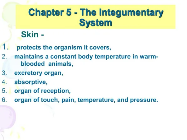
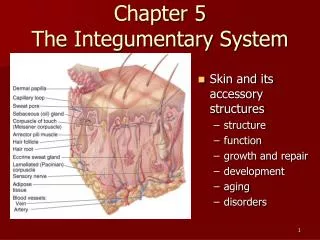
The Skin • It is the largest organ in the body, although it is easily infected because of its location.
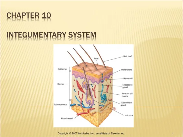
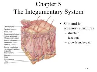
The Structure of Skin • Epidermis – superficial, thinner portion of epithelial tissue. • Dermis – intermediate, thicker, connective tissue layer. • Subcutaneous layer (hypodermis) – deep tissue layer that attaches skin to underlying structures.
The Function of Skin • Protection – outermost wall of defense • Physical barrier against; microbles, abrasions, dehydration, UV radiation (aids in production of Vitamin D) • Immunity • Temperature regulation • Regulates sweat secretions • Regulates blood flow close to the skin • Sensation • Skins acts as a big sensory organ (temp., pain, pressure) • Keeps body informed of changes in ethe envrionment
Epidermis • Its avascular • Types of cells • Keratinocyte – cells that produce keratin (protein) • Keratinization – process of filling skin cells with keratin as they migrate to the surface, which helps the skin as these cells reach the surface. The process takes about 2-4 weeks. • Melanocyte (also found in dermis) – cells that produce melanin – pigment used to color skin, eyes and hair.
Epidermis Continued • Langerhans cells- part of the immune response; easily damaged by UV rays • Merkel cell (found in the deepest layer of hairless skin) – thought to function in sensation of touch.
Epidermis Layers • Stratum basale (deepest layer) • Single layer • Anchors epidermis to basement membrane • Keratinization begin here • Cells are rapidly • Cells are rapidly dividing • Stratum spinosum • 8 to 10 layers • Keratin start to fibers accumulate within the cells
Epidermis Layers • Stratum granulosum • 2 to 5 layers thick • Cells are becoming flattened • In upper layers the cells die • Stratum lucidum • Cells are clear, flat and dead • Layer present in only a few areas in the body (thick & thin skin areas)
Epidermis Layers • Stratum corneum • 30 layers of dead cells • Cells are completely full of keratin • As new cells ascend old cells slough off
Dermis - vascular • Both the upper and lower regions contain connective tissue • Function – gives the skin strength • Characteristics – • Extensibility – ability to stretch • Elasticity – ability to return to its original shape, when skin can’t return to its normal shape stretch marks occur
Dermis • Structure- • Upper region (papillary) – areolar connective tissue • Dermal papillae – projections that increase surface area. • Cause ridges in epidermis • These ridges make our fingerprints • Meissner’s corpuscles – in some of the dermal papillae • Contain nerve endings • Contain blood capillaries
Dermis • Deep region (Reticular) • Contains • Dense irregular connective tissue (CT) • Adipose CT • Hair • Nerves • Oil glands • Sweat glands and duct • Heat and cold receptors
Dermis • Subcutaneous layer (hypodermis) • Function • Attaches dermis to underlying bone and muscle • Supplies dermis with blood vessels and nerves • Stores body fat (approx. ½ of the body’s fat)
Accessory Organs - Hair • Hair • Function – Protection from foreign particles and injury, cleanliness • Structure – • Shaft – superficial portion above the surface • Root – portion below the surface • Follicle – surrounds root composed of 2 layers • Bulb – onion shaped structure that surrounds the follice. • Matrix – where the hair is produced • Growth – 2 stages; resting and growth
Accessory Organs -Glands • Oil (sebaceous) • located by hair root • Secrets sebum(oil) • Function • Keeps hair from drying out • Prevents excess evaporation • Keeps skin soft • Cause blackheads and pimples • Gland becomes enlarged because a build up of waste products from the bacteria on the surface of the skin
Accessory Organs - Sweat • Sweat (sudoriferious) • Two types • Apocrine – found in armpit, pubic and areola • Open into hair follicles • Begin to function at puberty • Cause body odor when broken down by bacteria on the skin • Eccrine – found everywhere but in margin of lips, nail beds, and eardums • Most numerous in palms and soles • Function throughout life • normal
Accessory Organs - Wax • Wax (ceruminous) – located in outer ear canal • Produces cerumen • Secretions provide defense against foreign bodies
Accessory Organs - Nails • Structure – tightly packed keratinized cells • Nail body – visible portion • Free edge – extends past the tip of the finger • Nail root – not visible • Lunula – whitish cresent shape at base of the nail body • Matrix where the nail is produced • Function • Grasp and manipulate objects