Download
1 / 19
190 likes | 193 Views
Observing and measuring cells using a digital microscope. National 5 Cell Biology Cell structure Suggested learning activity.

E N D
Observing and measuring cells using a digital microscope.........
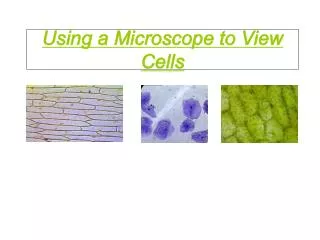
National 5 Cell Biology • Cell structure • Suggested learning activity Examine fresh and prepared slides of a range of plant, animal and microbial cells using appropriate stains and a light microscope/ bioviewer eg cheek epithelium, onion epidermis, rhubarb epidermis, Elodea, yeast. Numeracy activities on cell size to investigate cell length and breadth. Here we suggest using the Veho digital microscope to observe cells and to carry out measuring activities. TM
Start microscope • Connect microscope to PC USB port • Click icon on desktop • Choose an area, or object to view • Focus the microscope
Photo capture ‘thumbnails’ appear Mixed algae culture x 200
Some things to try • Photograph images of some of the following, noting the magnification • each time. • A coin / paper etc. • A pre-prepared microscope slide. • A slide of red onion tissue on a small drop of water. • A drop of saturated copper sulphate on a slide. Or use the Veho ‘lens cap’. Observe • crystals appearing. • Salt / sugar crystals. • Examine a prepared culture of algae. Use the Veho ‘lens cap’ as • a receptacle; clean the cap with alcohol and lens tissue. Keep the layer of • fluid very shallow.
Choose one, or two of your images and make some measurements: • Open the captured photo by double clicking on the thumbnail. • A preview window will open. • Input the magnification you noted for the image in the box at the top • right corner of the preview window. • You can measure the size of the whole or part of an object by using • the available options – direct line, multi-line, radius, etc.
units magnification direct line Sugar crystals x 200
x40 Accuracy and / or Precision? 0.90 mm 4.47 mm
Captured images can be annotated, or labelled. Don’t forget to save!