Download
1 / 21
270 likes | 835 Views
Ch 3: Observing Microorganisms Through a Microscope. Objectives Review the metric units of measurement Define total magnification and resolution Explain how electron and light microscopy differ Differentiate between acidic and basic dyes

E N D
Ch 3: Observing Microorganisms Through a Microscope
Objectives Review the metric units of measurement Define total magnification and resolution Explain how electron and light microscopy differ Differentiate between acidic and basic dyes Compare simple, differential, negative, and special stains List the steps in preparing a Gram stain. Describe the appearance of Gram-positive and Gram-negative cells after each step Compare and contrast Gram stain and acid-fast stain Explain why endospore and capsule stains are used
Units of Measurement Review Table 3.1 • 1 µm = ______ m = ______ mm • 1 nm = ______ m = ______ mm • 1000 nm = ______ µm • 0.001 µm = ______ nm
Sizes Among Microorganisms Compare to Foundation Fig 3.2 • Protozoa: 100 µm • Yeasts: 8 µm • Bacteria: 1 - 5 µm (some much longer than wide) • Rickettsia: 0.4 µm = _________ nm • Chlamydia and Mycoplasma: 0.25 µm • Viruses:20 – 250 nm Cells Alive – How big is a . . .?
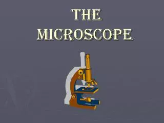
Principles of the Compound Light Microscope Magnification: Ocular and objective lenses of compound microscope (total mag.?) Resolution: Ability of lens to . . . Maximum resolving power depends on . . .For light microscope: ___ m Contrast: Stains change refractive index contrast between bacteria and surrounding medium Fig 3.1
RefractiveIndex • Measures light-bending ability of a medium • Light may bend in air so much that it misses the small high-magnification lens. • Immersion oil is used to keep light from bending. Fig 3.3
Brightfield Microscopy Simplest of all the optical microscopy illumination. techniques Dark objects are visible against a bright background. Darkfield Microscopy Light objects visible against dark background. used to enhance the contrast in unstained samples. Instrument of choice for spirochetes Microscopy: The Instruments Fig 3.4
Spirochetes (Treponema pallidum) viewed with darkfield microscope
Fluorescence Microscopy Uses UV light. Fluorescent substances absorb UV light and emit visible light. Cells may be stained with fluorescent chemicals (fluorochromes). Immunofluorescence Fig 3.6; T. pallidum
Fig 3.6 Principle of Immunofluorescence Figure 3.6a
Electron Microscopy:Detailed Images of Cell Parts Uses electrons, electromagnetic lenses, and fluorescent screens Electron wavelength ~ 100,000 x smaller than visible light wavelength Specimens may be stained with heavy metal salts Two types of EMs:?
SEM or TEM? Bacterial division Compare to Fig 3.10 Leaf surface
Preparation of Specimens for Light Microscopy • Staining Techniques Provide Contrast • Smear air-dry heat-fix • Basic dyes: cationic chromophore • Acidic dyes: anionic chromophore negative staining (good for capsules) • Three types of staining techniques: Simple, differential, and special
Use a single basic dye. A mordant may be used to hold the stain or coat the specimen to enlarge it. React differently with different bacteria Gram stain Acid fast stain Simple Stains Differential Stains
Figure 3.12 Gram staining. Gram-positive Gram-negative Application of crystal violet (purple dye) Application of iodine (mordant) Alcohol wash (decolorization) Application of safranin (counterstain) Rod (gram-negative) Cocci (gram-positive) Fig 3.12
Compare to Fig 3.12 Gram Stain crystal violet safranin
Gram Stains using Compound Light Microscope Streptococcus mutans Bacillus anthracis
Negative Stain Observe cell shape and size Used for bacteria with capsules Fig 3.14
Acid Fast Stain • Cells that retain a basic stain in the presence of acid-alcohol are called acid-fast. • Non–acid-fast cells lose the primary stain when rinsed with acid-alcohol, and are counterstained with a different color basic stain Fig 3.13
See Fig 3.14 Special Stains • Endospore stain: Heat is required to drive a stain into the endospore. • Flagella staining: requires a mordant to make the flagella wide enough to see. • Capsule stain uses basic stain and negative stain