Download
1 / 16
170 likes | 372 Views
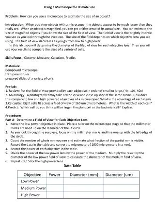
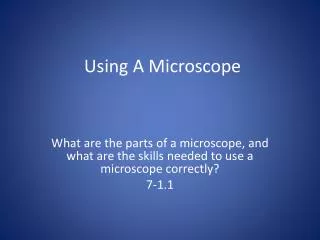
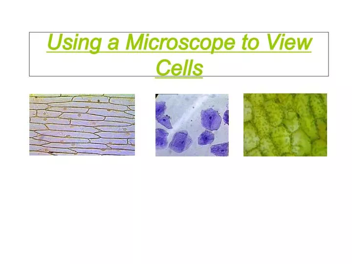
Using a Microscope to View Cells. Learning Targets. I can follow a lab procedure I can use prior knowledge, pictures and research to make connection and predictions between the investigation of the lab and my own experiences. I can use a microscope to view a microscopic image

E N D
Learning Targets • I can follow a lab procedure • I can use prior knowledge, pictures and research to make connection and predictions • between the investigation of the lab and my own experiences. • I can use a microscope to view a microscopic image • I can draw or take a picture a microscopic image using a camera and label the parts of the cell at 40X and 100X magnification using a compound light microscope. • I can distinguish difference between animal and plant cell based on shape, and characteristic organelle differences.
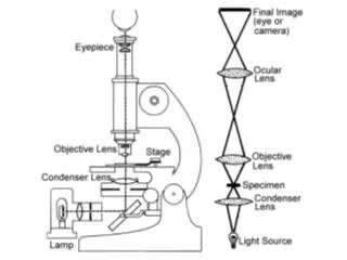
Microscope Procedure and Technique • Today we will make slides of 3 different cells and look at them under the microscope: • 1. Onion skin cells • 2. Elodea leaf cells • 3. Human cheek cells • Microscope rules • 1. Always carry or move a microscope with two hands, one on the arm, and one on the bottom. • 2. Always use the lowest power lens (the shortest lens) when you take a slide on and off the stage. • 3. Always start with the lowest power lens (the shortest lens). Get the slide in focus there, first using the coarse focus knob (the large knob) to get it close, THEN using the fine focus knob (the small knob) to get it perfectly in focus. From there, you can switch to a higher power lens. • 4. Always look from the SIDE of the microscope, not through the eyepiece, when switching lenses to avoid hitting the lens on the slide. • 5. Only use the coarse focus knob (the large knob) when you are using the lowest power lens (the shortest lens). Using the coarse focus knob on a higher power can crack the lens! • 6. Please turn off the light and cover the microscope when you are finished.
Preparing the Onion Cell Slide • Onion skin cells 1. Add 2 drops of iodine to the center of a glass slide. Be careful! Iodine can stain your clothes. 2. Take a small piece of onion. Use tweezers to peel off the skin from the underside (the rough, white side) of the onion. Throw the rest of the onion piece away. 3. Carefully lay the onion skin flat in the center of the slide on top of the iodine. 4. Add 2 drops of iodine to the top of the onion skin. 5. Stand a thin glass cover slip on its edge near the onion skin, next to the drop of iodine. 6. Slowly lower the other side of the cover slip until it covers the onion skin completely. If there are air bubbles, gently tap on the glass to “chase” them out.
Viewing the Onion Cells Under Low Power (40X) • 1. Make sure the lowest power lens (the shortest lens) is in place over the stage and the microscope light is turned on. Place the slide onto the stage of the microscope. • 2. Look through the eyepiece and turn the coarse focus knob (the largest knob) until an image comes into focus. It should look like a brick wall or like lizard skin. • 3. Now use the fine focus knob (the smallest knob) to make the image as focused as possible. • 4. Take a picture or draw what you see and insert it below.
Viewing Onion Cells Under Medium Power (100X) • 1. Again, looking from the SIDE of the microscope, rotate the lenses to the medium powered lens (100x). If you need to, use the fine focus knob (the smallest knob) to get the image into focus. You may see a dark blob in the middle of each cell. • 2. Try and capture the image with a camera • 3. Display and labeled the image below. • 2.In your lab notebook, draw a picture of what you see. Label the picture “Onion skin cells 400x”. Label as many parts of the cell as you can see. • 3. Switch to the lowest power lens and THEN remove the slide. Set it aside for now.
Viewing the Onion Cells under High Power (400X) • Increase the power to 400X by turning to the largest objective lens. • Using only fine focus, do your best to focus the image • Draw or take a picture. • Insert and label the image below • Switch to the lowest power lens and THEN remove the slide. Set it aside for now.
Preparing the Elodea Leaf Slide Preparing the Elodea leaf Slide • 1. Tear off one small leaf from the elodea plants floating in the fish tank. • 2. Add one drop of tap water to the slide. • 3. Stand a thin glass cover slip on its edge near the leaf, next to the drop of water. • 4. Slowly lower the other side of the cover slip until it covers the leaf completely. Make sure there are no air bubbles. • 5. Make sure the lowest power lens (the shortest lens) is in place over the stage. Place the slide onto the stage of the microscope.
Viewing Elodea Cells Under Lower Power (40X) • 1. Make sure the lowest power lens (the shortest lens) is in place over the stage. Place the slide onto the stage of the microscope. • 2. Make sure the lowest power lens (the shortest lens) is in place over the stage. Place the slide onto the stage of the microscope. • 3. Look through the eyepiece and turn the coarse focus knob (the largest knob) until an image comes into focus. It should look like small green bricks or like lizard skin. • 4. Now use the fine focus knob (the smallest knob) to make the image as focused as possible. • 5. In your lab notebook, draw a picture of what you see or Take a photo of your drawing. Go on line and find an actual picture of elodea cells. Label the picture “Elodea leaf cells 40x”. Label as many parts of the cell as you can see. • Insert image Below
Viewing Elodea Cells Under Medium Power • 1.Looking from the SIDE of the microscope, rotate the lenses to the 100x lens. If you need to, use the fine focus knob (the smallest knob) to get the image into focus. You should be able to see lots of small green dots in each cell. • 2.Record an image of the cells by drawing or camera and insert it below.
Elodea at High Power (400X) • 1. Again, looking from the SIDE of the microscope, rotate the lenses to the 400x lens. If you need to, use the fine focus knob (the smallest knob) to get the image into focus. The little green dots should get larger. • 2. In your lab notebook, draw a picture of what you see or take a picture. Insert Your image below. Go on line and find an actual picture of elodea cells. Label the picture “Elodea leaf cells 400x”. Label as many parts of the cell as you can see. • 3. Switch to the lowest power lens and THEN remove the slide. Set it aside for now.
Preparing the Human Cheek Cell Slide Preparing The Slide for Human cheek cells • 1.Add one drop of methylene blue to the middle of a clean slide. Be careful! Methylene blue will stain your clothes and skin. • 2.Use the flat side of a toothpick to gently scratch the inside of your cheek. DO NOT GOUGE YOUR CHEEK - you don’t need chunks of skin and definitely don’t want to draw blood. • 3.Gently touch the toothpick to the drop of dye on the slide. Some of your cheek cells should drift off into the dye. • 4.Throw the toothpick away. • 5.Stand a thin glass cover slip on its edge near the drop of dye. • 6.Slowly lower the other side of the cover slip until it covers the dye completely. Make sure there are no air bubbles.
Viewing Cheek Cells Under Low Power (40X) • 1.Make sure the lowest power lens (the shortest lens) is in place over the stage. Place the slide onto the stage of the microscope. • 2.Look through the eyepiece and turn the coarse focus knob (the largest knob) until an image comes into focus. It should look like scattered blobs. Move the slide around until a nice cluster of blobs moves into the center of your image. • 3.Use the fine focus knob (the smallest knob) to make the image as focused as possible. • 4. In your lab notebook, draw a picture or take a picture of what you see. Take a photo of your drawing. Label the picture “Human cheek cells 40x”.
Viewing Cheek Cells Under Medium Power (100X) • 1. Looking from the SIDE of the microscope, NOT through the eyepiece, rotate the lenses to the 100x lens. If you need to, use the fine focus knob (the smallest knob) to get the image into focus. • 2. Draw or take a picture of the image and insert below.
Viewing Cheek Cells Under High Power (400X) • 1. Looking from the SIDE of the microscope, rotate the lenses to the 400x lens. If you need to, use the fine focus knob (the smallest knob) to get the image into focus. • 2. In your lab notebook, draw a picture of what you see or take photo Take a photo of your drawing. Insert the picture below. Go on line and find an actual picture of cheek cells. Label the picture “Human cheek cells 400x”. Label as many parts of the cell as you can see. • 3. Switch to the lowest power lens and THEN remove the slide.
Questions and Tasks • Describe the shape of an animal cell vs. That of a plant cells • What organelles were visible that plant cells have but animal cells do not. • Why did the elodea cells we looked at have chloroplast but the onion cells did not? • Label the images with their cell type and magnification based on your images.