Download
1 / 76
870 likes | 1.02k Views
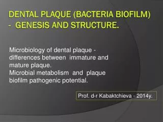
DENTAL PLAQUE. Most important chapter Understanding required Health maintenance Disease progression. OBJECTIVES. DEFINITION Difference between plaque, calculus , materia alba Types of plaque Composition of plaque Formation of plaque. Lecture 2. Microscopic structure of plaque

E N D
Most important chapter • Understanding required • Health maintenance • Disease progression
OBJECTIVES • DEFINITION • Difference between plaque, calculus , materia alba • Types of plaque • Composition of plaque • Formation of plaque
Lecture 2 • Microscopic structure of plaque • Concept of biofilm • Plaque Hypothesis • Microbial complexes • Microbial tests for plaque sample
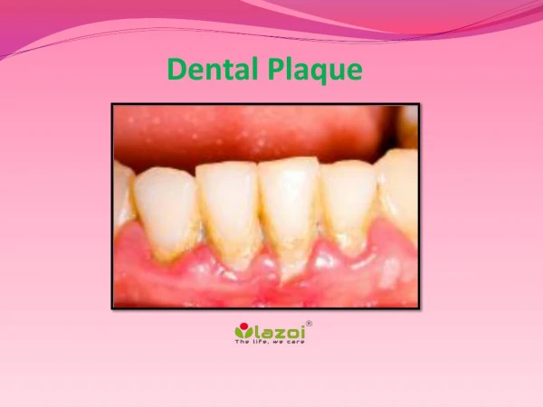
Definition • Dental Plaque “is a specific but highly variable structural entity, resulting from sequential colonization of microorganisms on tooth surfaces, restorations & other parts of oral cavity, composed of salivary components like mucin, desquamated epithelial cells, debris & microorganisms, all embedded in extracellular gelatinous matrix.” W.H.O ,1961
Dental Plaque can be defined as: “ soft deposits that form the biofilm adhering to the tooth surface or other hard surfaces in the oral cavity, including removable & fixed restorations” Bowen , 1976
Materia Alba “soft accumulations of bacteria and tissue cells; lack organised structure of dental plaque; easily displaced by water spray” • Dental Calculus ‘mineralised dental plaque”
DETECTION OF DENTAL PLAQUE DETECTION • Visual • Periodontal Probe or Explorer • Disclosing Agents
Timeline of plaque development: • At birth: sterile • Hours: facultative aerobic bacteria • Second day: anaerobic bacteria • 2 weeks: mature microbiota • Weaning (> 2 years): 400 different types of bacteria • Body contains 10 times more bacteria than human cells
Open growth system: communication with the pharynx • > 500 species in mouth • In any individual: 150 or more species at any given time.
Soft tissue Hard tissue Mastication Tongue Oral hygiene Washout effect: saliva Ciliae
Niches of plaque accumulation: • Supragingival and hard surgfaces: teeth, implant, restorations. • Periodontal pocket (hard: root cementum & soft: pocket epithelium) • Buccal, palatal and floor of the mouth epithelium • Dorsum of the tongue • Tonsils
Natural cleansing mechanism: • Gingival crevicular fluid & salivary flow • Cleansing effect of mastication and tongue movement • Rapid turnover rate of intraoral epithelial cells • Host defence mechanisms like langerhans cells.
Hard tissues • Teeth and implants surface provide a non shedding surface. It allows extensive plaque accumulation. • Provide an ectodermal interruption. At junctional epithelium the teeth provide access to bacteria into the body. • Port of entry of periopathogens.
Macroscopic Structure • Supragingival Plaque • Subgingival Plaque • Marginal Plaque
Composition of Plaque • Microorganisms • Intercellular Matrix
Microorganisms Bacterial 1gm contains 2X10¹¹ bacteria-500 species Non-Bacterial Mycoplasma, Yeast, Protozoa, Viruses
Intercellular Matrix • Accounts for 20% to 30% of the plaque mass • Organic and inorganic materials • Derived from saliva, gingival crevicular fluid, and bacterial products.
Organic Component • Organic constituents : polysaccharides, proteins, glycoproteins, and lipid material • Polysaccharides produced by bacteria-Dextran: predominant form • Albumin: originating from crevicular fluid • Lipid material: debris from the membranes of disrupted bacterial and host cells and possibly food debris.
Inorganic Component • Calcium and phosphorus (Most) • Trace amounts: sodium, potassium and fluoride. • Source -supragingival plaque (saliva) & subgingival plaque (GCF & Blood) • Calculus frequently found in areas of the dentition adjacent to salivary ducts
PHASES OF PLAQUE FORMATION
Formation of dental Pellicle • Initial Colonization • Secondary Colonization & Plaque Maturation
Formation of dental Pellicle • Acquired enamel pellicle forms rapidly - Early pellicle • Characterized by an absence of bacteria and their products. • Composed of proteins and glycoproteins. • Demonstrate a higher content of threonine, serine, and alanine & less proline than saliva- selective adsorption
Electron microscopic -reveals a thin, amorphous, electron-dense layer immediately adjacent to the hard surface- thickness from 1 to 2 nm. • involves a combination of physical forces (ionic, hydrophobic, hydrogen bonding, and van der Waals)
May involve the interaction of phosphate groups with calcium ions in saliva to form "bridges" • Protective functions of early enamel pellicle: protection , lubrication by decreasing frictional forces, may selectively concentrate antimicrobial substances such as immunoglobulins, lysozyme, and cystatins at different oral surfaces.
Formation of later pelliclemost likely involves protein-protein or protein-carbohydrate interactions,- stereospecific in nature . • For example, A. viscosus and Streptococcus mitis produce a neuraminidase that cleaves terminal sialic acid residues on the glycoproteins in saliva or early pellicles to expose galactose residues (Costello et aI., 1979) • Collectively, these mechanisms may be important for the initial colonization
Initial adhesion Phase I : Transport to the surface Phase II : Initial adhesion Phase III : Attachment Phase IV : Colonisation of the surface and biofilm formation
Phase I • Random contacts: • Brownian motion • Sedimentation of micro- organisms • Liquid flow • Active bacterial movement
Phase II • Initial reversible adhesion • Long range and short range forces: • van der Walls attractive forces • Electrostatic repulsive forces
Phase III • Firm anchorage • Specific interactions: • Covalent • Ionic • Hydrogen bonding
Adhesions: specific extracellular proteinacious components on microorganisms. • Complimentary receptors: proteins, glycoproteins or polysaccharides on the pellicle surface.
Example: • S. Sanguis- binds to acidic proline rich proteins, alpha amylase & sialic acid • A. viscosus- fimbriae that contain adhesins- bind to proline rich proteins of dental pellicle.
Colonisation & Plaque maturation • Initial colonisers attach to the tooth surface- provide substrate for secondary colonisers to attach. • They create a favourable environment for secondary colonisers to survive.
Secondary Colonisers: • Adhere to bacteria already present in the plaque mass. • Prevotella intermedia • Prevotella loeschii • Fusobacterium nucleatum • Porphyromonas gingivalis • Capnocytophaga
Co- aggregation • Cell to cell recognition of genetically distinct partner cell types • Highly specific stereochemical interaction • Corn cob formation: long filament bacteria covered with cocci • Test tube brush: long filament bacteria covered with flagellated small motile rods
MICROSCOPIC STRUCTURE SUPRAGINGIVAL PLAQUE • typically demonstrates a stratified organization of the bacterial morphotypes. • Gram-positive cocci and short rods predominate at the tooth surface • gram-negative rods and filaments ,spirochetes predominate in the outer surface of the mature plaque mass. • Highly specific cell-to-cell interactions are also evident from the "corncob"
CORN COB STRUCTURE CORN-COB STRUCTURE
Thin section of supragingival plaque GRAM POSITIVE BACTERIA IN PALISADING ARRANGEMENT
SUBGINGIVAL PLAQUE • Gingival crevicular fluid, -contains many substances that the bacteria may use as nutrients • Host inflammatory cells and mediators have influence on the establishment and growth of bacteria in this region. • Distinctions present between tooth-associated and tissue-associated regions of subgingival plaque
Thin section of plaque in a deep pocket RODS COCCI FILAMENTS
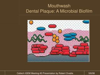
Plaque-A Biofilm 1.Structure-microcolonies of bacteria in matrix 2.Exopolysaccharides produced by bacteria 50-90% of dry wt integrity of biofilm buffer
3.Physiological Heterogenicity Same sp.-different physiologic states in biofilms 4.Quorum Sensing “regulation of expression of specific genes”- intercellular communication distinct properties Prosser,1999 5.Attachment of Bacteria- fimbrae & fibrils
6.Increased antibiotic Resistance Bacteria in biofilms more resistant Slow growth Decreased diffusion Accumulation of enzymes- lactamases,dehydrogenases Alteration of genes.