Download
1 / 97
1k likes | 1.59k Views
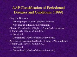
Etiology, pathogenesis, clinical manifestation, diagnosis of generalized periodontitis and periodontal disease. I) GINGIVAL DISEASE A) Dental plaque induced 1) Gingivitis associated with dental plaque only | 2) Gingival diseases modified by systemic factors

E N D
Etiology, pathogenesis, clinical manifestation, diagnosis of generalized periodontitis and periodontal disease
I) GINGIVAL DISEASE A) Dental plaque induced 1) Gingivitis associated with dental plaque only | 2) Gingival diseases modified by systemic factors Example: Bleeding on probing | a) Associated with endocrine system a) Without other local contributing factors | 1) Puberty b) With local contributing factors | 2) Menstrual cycle Example: Restorations | 3) Pregnancy Mouth breathing | Examples: a) Gingivitis | b) Pyogenic granuloma
I) GINGIVAL DISEASE (continued) A) Dental plaque induced 2) Gingival diseases modified by systemic factors a) Associated with endocrine system 4) Diabetes mellitus associated gingivitis Examples: I Role of diabetes in periodontal disease II Periodontal disease in diabetic patients. Increased risk of periodontal abscess, increased gingival reaction to plaque, increased risk of periodontal disease. b) Associated with blood dyscrasias 1) Leukemia-associated gingivitis - Examples: Bleeding into gingival tissue Gingival enlargements 2) Other
I) GINGIVAL DISEASE (continued) B) Non plaque induced gingival lesions 3) Gingival diseases of fungal origin | 5) Gingival manifestations of systemic conditions a) Candida species infections | a) Mucocutaneous disorders 1) Generalized gingival candidiasis | 1) Lichen planus b) Linear gingival erythema | 2)Pemphigoid Example: HIV associated gingivitis | 3) Pemphigus vulgaris AIDS related periodontitis | 4) Erythema multiforme c) Histoplasmosis | 5) Lupus erythematosus d) Other | 6) Drug induced 4) Gingival lesions of genetic origin | 7) Other a) Hereditary gingival fibromatosis | b) Other | | |
I) GINGIVAL DISEASE (continued) B) Non plaque induced gingival lesions 5) Gingival manifestations of systemic conditions b) Allergic reactions 1) Dental restorative materials | 3) Traumatic lesions (factitious, iatrogenic, a) Mercury | accidental) b) Nickel | a) Chemical injury c) Acrylic | Example: Hydrogen peroxide, d) Other - Example: Nickel allergy | aspirin burn 2) Reactions attributable to | b) Physical injury - Example: a) Tooth paste | toothbrush trauma, cotton roll burn b) Mouth rinse | c) Thermal injury c) Chewing gum | 4) Foreign body reactions d) Food and additives - Examples: | 5) Not otherwise specified Example: Gingival allergy to cinnamon | Cocaine induced gingival necrosis
II) CHRONIC PERIODONTITIS A) Localized Example: Molar furcation, premolar, intrabony defect B) Generalized Example: Upper molars and premolars III) AGGRESSIVE PERIODONTITIS A) Localized - Example: Juvenile onset periodontitis. Affects first molars and incisors with little signs of gingival inflammation. May be related to: a) Actinibacillus actinomycetemcomitans. B) Generalized IV) PERIODONTITIS AS MANIFESTATION OF SYSTEMIC DISEASE A) Associated with hematologic disorders 1) Acquired neutropenia 2) Leukemias 3) Other
IV) PERIODONTITIS AS MANIFESTATION OF SYSTEMIC DISEASE (continue) B) Associated with genetic disorders 1) Familial and cyclic neutropenia Example: ANUG type lesions that do not respond to local therapy. 2) Down syndrome a) See high prevalence of advanced periodontitis. 1 in 800 incidence. Chromosomal disorder e.g. Trisomy 21 (three chromosomes). More common in older mothers. 3) Leukocyte Adhesion Deficiency Syndromes b) Leukocytes can’t adhere to blood vessels and migrate to inflammatory sites. Get recurrent infection. 4) Papillon Lefévre syndrome Example: Aggressive periodontitis in children with hyperkeratotic lesions of hands, knees and feet. Autosomal recessive inheritance. Incidence 4 per million. 5) Chediak-Higashi syndrome c) Functional neutrophil defects of chemotasis and bacterial killing. See severe periodontitis
IV) PERIODONTITIS AS MANIFESTATION OF SYSTEMIC DISEASE (continue) B) Associated with genetic disorders 6) Histiocytosis syndrome d) Cause unknown. Increase in monocytes and macrophages. Lesions in bone and gingival swelling. 7) Glycogen storage disease e) Many types of genetics upsets,to enzymes with liver dysfunction. Incidence 1 in 25,000. 8) Infantile genetic agranulocytosis 9) Cohen syndrome f) Autosomal recessive, short head and upper lip exposure of incisors. 10) Ehlers-Danlos syndrome g) Group of inherited disorders of collagen, joint affected. Increased tissue fragility, poor healing. 11) Hypophosphatasia Example: Disturbance to bone metabolism, loss of primary teeth. Aggressive juvenile type periodontitis 12) Other C) Not otherwise specified
V) NECROTIZING PERIODONTAL DISEASES A) Necrotizing ulcerative gingivitis Example: Associated with large amounts of fusiforms and spirochetes. Mainly adults. Only affects children that have severe systemic problems like malnutrition. B) Necrotizing ulcerative periodontitis Example: Can be associated with AIDS VI) ABSCESSES OF THE PERIODONTIUM A) Gingival abscess Example: Localized to gingival tissue B) Periodontal abscess Example: Spread to involve larger area C) Pericoronal abscess VII) PERIODONTITIS ASSOCIATED WITH ENDODONTIC LESIONS A) Combined periodontic endodontic lesions Examples: Need to have radiologic evaluation and vitality testing
VIII) DEVELOPED OR ACQUIRED DEFORMITIES AND CONDITIONS A) Localized tooth related factors that modify or predispose to plaque induced gingival disease, periodontitis 1) Teeth anatomic factors Example: Development at groove on palatal of upper lateral incisor 2) Dental restorations Example: Over contoured crowns. Poorly fitting margins 3) Root fracture Example: Longitudinal fractures have hopeless prognoses B) Mucogingival deformities and conditions around teeth 1) Gingival soft tissue recession a) Facial or lingual surfaces Example: Inadequate band of keratinized gingiva b) Interproximal papillary Examples: Loss of anterior papilla
VIII) DEVELOPED OR ACQUIRED DEFORMITIES AND CONDITIONS (Continued) B) Mucogingival deformities and conditions around teeth 2) Lack of keratinized gingiva 3) Decreased vestibular depth 4) Aberrant frenum 5) Gingival excess a) Pseudopocket b) Inconsistent gingival margin c) Excessive gingival display Example: Poor gingival esthetics d) Gingival enlargement i) See 1A3, 1A4 e) Abnormal color
VIII) DEVELOPED OR ACQUIRED DEFORMITIES AND CONDITIONS (Continued) C) Mucogingival deformities and conditions on edentulous ridges 1) Vertical and/or horizontal ridge deformity Example: Ridge deformities 2) Lack of gingiva keratinized tissue 3) Gingival/soft tissue enlargements 4) Decreased vestibular depth 5) Abnormal color D) Occlusal Trauma 1) Primary occlusal trauma 2) Secondary occlusal trauma
I) Gingival disease A) Dental plaque induced 1) Gingivitis associated with dental plaque only Example: BLEEDING ON PROBING One of the earliest signs of gingivitis is bleeding on probing.
I) Gingival Disease (Continued) A) Dental plaque induced 1) Gingivitis associated with dental plaque only b) With local contributing factors Example: RESTORATIONS Inflammation with pocket depth restricted to gingival tissues.
I) Gingival Disease (Continued) A) Dental plaque induced 1) Gingivitis associated with dental plaque only b) With local contributing factors Example: MOUTH BREATHING This type of gingivitis affects the anterior gingiva of chronic mouth breathers or individuals with incomplete lip closure. Note the erythematous, hypertrophic maxillary anterior gingiva.
Gingival Disease (Continued) A) Dental plaque induced 2) Gingival diseases modified by systemic factors a) Associated with endocrine system 1) Puberty 2) Menstrual cycle 3) Pregnancy Examples: a) Gingivitis b) Pyogenic granuloma The gingival tissues may have a modified reaction to dental plaque with changes in circulating estrogen and progesterone levels. These changes result in the inflammation having more vascular components and this is generally not very obvious in puberty or with menstrual cycles but can be quite pronounced in some pregnant patients.
I) Gingival Disease (Continued) A) Dental plaque induced 2) Gingival diseases modified by systemic factors a) Associated with endocrine system 3) PREGNANCY GINGIVITIS These are two examples of pregnancy gingivitis. Note the intense burgundy color and the marked gingival hypertrophy. These lesions bleed profusely.
I) Gingival Disease (Continued) A) Dental plaque induced 2) Gingival diseases modified by systemic factors a) Associated with endocrine system 3) PYOGENIC GRANULOMA Pyogenic granuloma is considered to be a exuberant response to a chronic mild irritant. Its clinical appearance is similar to that seen in pregnancy gingivitis but generally confined to a single area. Pyogenic granulomas also bleed easily because they contain multiple capillaries.
For details click on the books Gingival disease (Continued) A) Dental plaque induced 2) Gingival disease modified by systemic factors a) Associated with endocrine system 4) DIABETES MELLITUS ASSOCIATED GINGIVITIS Note the marked inflammatory reaction and hypertrophy of the free gingiva in this patient with diabetes mellitus. This reflects an increased gingival reaction to plaque with consequent increased risk of periodontal disease.
Periodontal disease in diabetic patients increased incidence of periodontal abscesses increase gingival inflammatory reaction to plaque increase risk of periodontal disease 2.8 to 3.4 increase increase severity and rate of destruction. Attachment and bone loss twice as much in diabetic Pima Indians compared with controls
Role of Diabetes in Periodontal disease Reduce vasculature efficiency PMN defects Macrophage increase cytokines with P. Gingivalis 24 to 32 times more TNF 4 times increase in PGE and ILI Increase collagenase Increase in cross linked collagen by AGEs. Delayed healing and repair
I) Gingival disease (Continued) A) Dental plaque induced 2) Gingival disease modified by systemic factors a) Associated with endocrine system 4) DIABETES MELLITUS PERIODONTAL ABSCESS There is a greater increase risk for diabetic patients to develop periodontal abscesses due to increased gingival reaction to plaque and increased risk of periodontal disease. The arrow points to the abscess.
Poor diabetic control and length of time increase risk of periodontal breakdown and increase chances of poor response to therapy.
I) Gingival disease (Continued) A) Dental plaque induced 2) Gingival disease modified by systemic factors b) Associated with blood dyscrasias 1) LEUKEMIA ASSOCIATED GINGIVITIS For details click on the books Note the generalized facial pallor and skin echymosis. The gingiva is hypertrophic and shows a typical intragingival hemorrhage.
For details click on the books I) Gingival disease (Continued) A) Dental plaque induced 3) Gingival diseases modified by medications a) Drug induced gingival disease 1) PHENYTOIN GINGIVAL HYPERTHROPHY Phenytoin gingival hypertrophy has an incidence of 3 to 84.5%. This enlargement is produced by hyperplasia of the connective and epithelial tissues with secondary inflammation. It may have increased expression of platelet derived growth factor.
CALCIUM CHANNEL BLOCKERS OF SMOOTH AND CARDIAC MUSCLE ° TRADE NAME VERAPAMIC CALAN DILTIAZEM CARDIAZEM FECODIPINE PLENDIL ESRAPIDINE PRESCAL NICARDIPIDINE CARDENE NIFEDIPIDINE PROCARDIA NISOLPIDINE SYSLOC NITRENDIPIDINE BAYOTENSIN MIMODIPIDINE NIMOTOP
For details click on the books I) Gingival disease (Continued) A) Dental plaque induced 3) Gingival diseases modified by medications a) Drug induced gingival disease 1) CALCIUM CHANNEL BLOCKERS - NIFEDIPINE Nifedipine is used for coronary artery disease and hypertension to dilate blood vessel and is also used with immunosuppressant drugs in organ transplant. This medication induces gingival hypertrophy, as seen here, in 25% to 50% of patients.
I) Gingival disease (Continued) A) Dental plaque induced 3) Gingival diseases modified by medications a) Drug induced gingival disease 1) IMMUNOSUPPRESANT CYCLOSPORINE For details click on the books Cyclosporin A is an immunosuppressant used in organ transplant and it produces gingival enlargement in at least 30% of patients under treatment.
I) Gingival disease (Continued) A) Dental plaque induced 3) Gingival diseases modified by malnutrition a) ASCORBIC ACID GINGIVITIS For details click on the books This gingivitis seen only in the late stages of scurvy is plaque associated. Severe vitamin C deficiency induces absence of intracellular oxidation, abnormal collagen formation, gingival hypertrophy with hemorrhage and mucosal echymoses.
I) Gingival disease (Continued) B) Non plaque induced 1) Gingival diseases of specific bacterial origin Example: RECURRENT APHTOUS STOMATITIS For details click on the books Recurrent aphtous stomatitis is divided in aphthous minor, aphthous major and herpetiform ulcers. Aphthous minor rarely affects the gingiva. These ulcers are very painful and may last up to 14 days.Etiolgy is unknown.
Gingival disease (Continued) B) Non plaque induced 2) Gingival diseases of viral origin a) Herpes virus - PRIMARY HERPETIC GINGIVOSTOMATITIS For details click on the books To the left a 13 y.old boy and to the right a 23 y.old man both with primary herpetic gingivostomatitis. Note gingival bleeding and ulcerations which were preceded by vesicles. Also note sero-purulent exudate in the 23 y.old man.
Gingival disease (Continued) B) Non plaque induced 2) Gingival diseases of viral origin a) Herpes virus - RECURRENT INTRAORAL HERPES SIMPLEX For details click on the books The intraoral lesions of RHS are characterized by small linear vesicles that rupture and leave small areas of ulceration. Both the free and attached gingiva can be the site of these lesions.
Gingival disease (Continued) B) Non plaque induced 2) Gingival diseases of viral origin a) Herpes virus - RECURRENT INTRAORAL HERPES SIMPLEX GINGIVAL MUCOSAL LESIONS These intraoral recurrent lesions of herpes simplex resulted from the minor trauma associated with root planing. Note the marked involvement one week after root planing.These lesions are infrequently seen and may occur after flap surgery.
I) Gingival disease (Continued) B) Non plaque induced 2) Gingival diseases of viral origin a) Herpes virus - HERPES ZOSTER INFECTION For details click on the books Skin and mucosal lesions of herpes zoster are characterized by linear crops of vesicles, as seen here. When the intraoral vesicles break leave painful ulcers. Post zoster neuralgia is a frequent sequela.
I) Gingival disease (Continued) B) Non plaque induced 2) Gingival diseases of viral origin a) Herpes virus - HERPES ZOSTER INFECTION Herpes Zoster lesions follow the affected nerve distribution,in this case the Mandibular branch of the Trigeminal nerve. To the right healing 3 weeks later.
I) Gingival disease (Continued) B) Non plaque induced 2) Gingival diseases of viral origin a) Herpes virus - AIDS RELATED KAPOSI SARCOMA For details click on the books These are two examples of gingival Kaposi sarcoma. To the left generalized gingival involvement . To the right a localized sarcoma mimicking a pyogenic granuloma. Herpes virus 8 is considered the etiologic agent of AIDS related Kaposi sarcoma.
Gingival disease (Continued) B) Non plaque induced 3) Gingival diseases of fungal origin a) Candida species infections 1) GENERALIZED GINGIVAL CANDIDIASIS For details click on the books The left is an example of acute pseudo- membranous candidiasis (thrush),white lesions that can be lifted off the gingiva.The other case to the right shows an example of acute atrophic (eythematous) gingival candidiasis.
ORAL MANIFESTATIONS OF AIDS|AIDS and the PERIODONTIUM ° Hairy leukoplakia |° Linear gingival erythema ° Candidiasis |°Necrotizing ulcerative periodontitis ° Other mycotic infections |° Necrotizing stomatitis ° Oral ulcers and delayed healing |° Candidiasis ° Herpetic infections |° Other mycotic infections ° Other viral infections |° Herpetic infections ° Kaposi’s sarcoma |° Other viral infections ° Other lesions |° Kaposi’s sarcoma
For details click on the books I) Gingival disease (Continued) B) Non plaque induced 3) Gingival diseases of fungal origin b) Linear gingival erythema HIV ASSOCIATED GINGIVITIS Note the well delineated erythematous band following the contour of the free gingival margin. This phenomenon reflects inflammation as a consequence to bacterial invasion and proliferation in the gingival sulcus.
Gingival disease (Continued) B) Non plaque induced 3) Gingival diseases of fungal origin b) Linear gingival erythema AIDS RELATED PERIODONTITIS For details click on the books The photo to the left shows areas of gingival and periodontal necrosis and gingival hypertrophy. The photo to the right shows marked gingival recession and bone exposure.These lesions can destroy tissue rapidly Both patients were HIV positive.
For details click on the books Gingival disease (Continued) B) Non plaque induced 4) Gingival lesions of genetic origin a) Hereditary gingival fibromatosis AUTOSOMAL DOMINANT GINGIVAL FIBROMATOSIS Marked gingival hypertrophy in a patient with autosomal dominant gingival fibromatosis.This is seen early affecting even the deciduous dentition. The teeth are partially covered and eruption is retarded.
Gingival disease (Continued) B) Non plaque induced 4) Gingival lesions of genetic origin b) Other This patient is an example of a syndrome characterized by gingival hyperplasia, increased growth of hair, epilepsy and mental retardation, inherited as an autosomal dominant. Note the increased amount of facial hair and the gingival fibromatosis.
Note the striations and erosion of the gingiva. Lichen planus may be an autoimmune response. Vesicles may be present, lace like white lesions of gingiva, tongue and cheek are also part of the clinical manifestations. In some patients the ulcerations may be related to friction. For details click on the books I) Gingival disease (Continued) B) Non plaque induced gingival lesions 5) Gingival manifestations of systemic conditions a) Mucocutaneous disorders - LICHEN PLANUS
I) Gingival disease (Continued) B) Non plaque induced gingival lesions 5) Gingival manifestations of systemic conditions a) Mucocutaneous disorders - LICHEN PLANUS These are examples of squamous cell carcinoma arising in a previous erosive Lichen Planus observed in two different patients.There may be an increased risk of neoplastic change in Lichen Planus.
I) Gingival disease (Continued) B) Non plaque induced gingival lesions 5) Gingival manifestations of systemic conditions a) Mucocutaneous disorders - MUCOUS MEMBRANE PEMPHIGOID For details click on the books These photos show gingival erythema and desquamation with symptons of gingival pain in two patients with Benign Mucous Membrane Pemphigoid.
I) Gingival disease (Continued) B) Non plaque induced gingival lesions 5) Gingival manifestations of systemic conditions a) Mucocutaneous disorders - MUCOUS MEMBRANE PEMPHIGOID The drawing and the microscopy show the vesicle formation beginning at the Basement Membrane typical of Benign Mucous Membrane Pemphigoid.
I) Gingival disease (Continued) B) Non plaque induced gingival lesions 5) Gingival manifestations of systemic conditions a) Mucocutaneous disorders - MUCOUS MEMBRANE PEMPHIGOID Indirect immunofluorescence shows that an antibody-antigen reaction is present at the level of the epithelial basement membrane as an auto immune response.
For details click on the books I) Gingival disease (Continued) B) Non plaque induced gingival lesions 5) Gingival manifestations of systemic conditions a) Mucocutaneous disorders - PEMPHIGUS VULGARIS These photos of the same patient show gingival desquamation, ulcers, erythema and vesicle formation. These were the initial painful manifestations of Pemphigus in this patient.
I) Gingival disease (Continued) B) Non plaque induced gingival lesions 5) Gingival manifestations of systemic conditions a) Mucocutaneous disorders - PEMPHIGUS VULGARIS The drawing and the microscopy demonstrate the intraepithelial vesicle formation typical of Pemphigus Vulgaris. Also note Tzank cells within the vesicle lumen.
I) Gingival disease (Continued) B) Non plaque induced gingival lesions 5) Gingival manifestations of systemic conditions a) Mucocutaneous disorders - PEMPHIGUS VULGARIS Direct immunofluorescence of Pemphigus Vulgaris shows that the auto immune antibody-antigen reaction is present within the gingival epithelial intercellular adhesion system. This affects the desmosomes of the spinal cell layer. The result is acantholysis, that is cellular detachment and vescicles.