Download
1 / 95
1.04k likes | 1.25k Views
DENTAL PLAQUE. Contents. Introduction Definition and structure Composition Classification Stages of plaque formation Plaque as a biofilm. Human fetus →sterile oral cavity. Hours after birth→Facultative aerobic bacteria. Second day→anaerobic bacteria .

E N D
Contents • Introduction • Definition and structure • Composition • Classification • Stages of plaque formation • Plaque as a biofilm
Human fetus →sterile oral cavity. Hours after birth→Facultative aerobic bacteria. Second day→anaerobic bacteria . Two weeks→nearly mature microbiota in the gut
Oral cavity - >500 bacterial species, mostly commensal & beneficial • Open growth system • periodontal diseases unusual human infection. major reason anatomic feature of a mineralized structure, the tooth, partly exposed to the external environment and partly in the connective tissues
Teeth & implants – hard non shedding surfaces, continually held in immediate proximity to the soft tissues of the periodontium • Provide port of entry
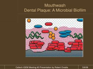
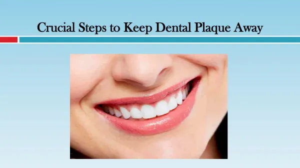
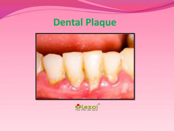
Dental Plaque - Definitions • “ A soft deposit that form the biofilm adhering to the tooth surfaces in the oral cavity including removable and fixed restorations. • Diverse community of microorganism found on tooth surface as biofilm embeded in an extracellular matrix of polymers of host and microbialorigin (Marsh 2004)
Clinically plaque is def as structured resilient, yellow grayish substance that adheres tenaciously to the intraoral hard surfaces including removable and fixed restorations • Accumulates in the gingival third and in pits, cracks, fissures, overhanging restorations and around malaligned tooth
Methods of detecting plaque • Probing with a periodontal probe. • Disclosing solution.
Materia Alba - Soft accumulations of bacteria and tissue cells that lack the organised structure of dental plaque and are easily displaced by water spray. • Calculus – Hard deposit that forms by mineralisation of dental plaque and is generally covered by a layer of unmineralised plaque
Composition • Water: 80-85% plaque mass ; • Cells: primarily bacteria, 1 gm (wet weight)= 1011 bacteria • Non bacterial: Mycoplasma spp, yeasts, protozoa, viruses • (conteras 2000) • Host cells : epithelial cells, macrophages & leucocytes • Matrix: Organic Inorganic
Matrix : 20-30% plaque mass • Organic: Carbohydrates: Dextrans, levans, polysaccharides, galactose Lipids Proteins: Albumin, glycoproteins Misc: cxtracellular bacterial products, cell remnants, food • Inorganic: Calcium Phosphorus/ phosphate Na, Cl, F
SUPRAGINGIVAL PLAQUE • Gram –positive cocci and short rods predominate at the tooth surface area, • Gram –ve rods filaments as well as spirochaetes pedominate the outer surface of mature plaque mass.
TOOTH ASSOCIATED ( Attached Plaque) • Cervical part : densely arranged consists of gram +ve rods and cocci including Streptococcus mitis , Streptococcus sanguis, Actinobacillus Viscosus, A. naeslundi and Eubacterium species . • Deeper parts filamentous forms are fewer • Apical border of plaque mass is separated from junctional epithelium by a layer of host leukocytes. increased concentration of gramnegative rods, spirochetes
TISSUE / EPITHELIUM ASSOCIATED PLAQUE -Loosely arranged - Comprises of Gram-negative rods and cocci, large number of filaments, flagellated rods, spirochaetes. Presence of Porphyromonas gingivalis, Prevotella Intermedia, Tannerella Forsythia , Fusobacterium Nucleatum are predominant -
Clinical signifance • Marginal plaque→ gingivitis • Supragingival and tooth associated subgingival plaque→calculus formation • Tissue associated subgingival plaque→periodontal tissue destruction • Subgingival tooth attached plaque→calculus formation and root caries
Plaque formation 3 major phases of plaque formation
MECHANISMS OF PLAQUE FORMATION • Formation of pellicle • Pellicle – Glycoprotein derived from components of saliva and GCF as well as bacterial and host tissue cell products and debris. • Forms within nanoseconds after vigorous brushing • Contains – glycoproteins (mucins), Phosphoproteins, Histidine rich proteins, enzymes (α- amylase) • Forms by selective adsorption of the environmentalmacromolecules • mechanisms involved in enamel pellicle formation include electrostatic, van der Waals, and hydrophobic forces
. • Can be removed by polishing . • Recurs soon after removal. • Fully established pellicle – in 30 minutes.
Initial Adhesion and Attachment of Bacteria Phase I: Transport to the surface • Random contacts through brownian motion. • Sedimentation of microorganisms • Through liquid flow • Active bacterial movement ( chemotactic activity).
Phase 2 ; Initial adhesion • Reversible adhesion of bacterium to the surface • Through long and short range forces like van der Waalsattractive and elecrostatic repulsive forces, hydrogen bonding
Phase 3 : Attachment Firm anchorage→ specific interactions ( covalent, hydrogen bonding ) Direct contact or bridging through specific extracellular proteinecious components of organisms and complementory receptors ( proteins and glycoproteins or polysaccharides on the surface )
S. sanguis ( principle early coloniser )bind to proline rich proteins • A. viscosus posses fimbriae →adhesins →proline rich proteins . .
Colonisation and plaque maturation • Plaque increases by two mechanisms. • Multiplication of bacteria already attached to the tooth surface. • Subsequent attachment and multiplication of new bacterial species to cells of bacteria already present in the plaque mass.
Coaggregation • Coaggregation - defined as the specific cell-to-cell recognition that occurs between genetically distinct cell types • Occurs primarily through highly specific stereochemical interaction of protein and carbohydrate molecules located on the bacterial cell surface. • Less specific interactions from hydrophobic, electrostatic and vanderWaals forces also occur.
Early stages, coaggregation occurs among different gram positive species and between gram positive and gram negative species. Later stages between different gram negative species . Eg: E.nucleatum,P. gingivalis and T. denticola
Primary colonizers are taught to prepare a favorable environment for secondary colonizers They are gram +ve aerobic micro organisms • secondary colonizers do not initially colonize on to the clean tooth surface but adhere to bacteria already in the plaque mass They are gram -ve anaerobic micro organisms
Secondary colonizers • Gram negative species- Fusobacterium nucleatum, Prevotella intermedia , Porphyromonas gingivalis, Prevotella loeschii and capnocytophaga species. • Adehere to the gram positive species already present in the plaque..
Tertiary colonisers After one week of plaque accumulation other gram negative species may also appear P gingivalis, C rectus., E corrodens A A Comitans and the oral spirochaetes( Treponema species)
Spatiotemporal model of Oral bacterial colonization Kolenbrander et al Perio 2000 vol-42
Socransky etal →13000 plaque samples Used cluster analysis and community coordination techniques to demonstrate the presence of specific microbial groups. DNA hybridisation methodology defined “ complexes” of periodontal microorganisms
Yellow complexes -streptococcus species. • Purple complex consisting of Veillonella parvula and Actinomyces odontolyticus. • Green Complex: E corrodens, A. actinomycetemcomitans serotype a, and Capnocytophaga species. • Orange Complex : Fusobacterium , prevotela and Campylobacter species. • Red Complex:P gingivalis, Tanerella forsythus and T denticola. They are aasociated with bleeding on probing.
Microbial Complexes Actinomyces species SUBJECTS - 185 PLAQUE SAMPLES- 13,261 40 subgingival microorganisms DNA-hybridization Socransky et al - 1998 V.parvula A.odontolyticus S.mitis S.Sanguis S.oralis P.intermedia P.nigrescens P.micros F.nucleatum C.rectus P.gingivalis T.forsythia T.denticola E.corrodens capnocytophaga A.a BOP
Physiological properties of dental plaque • Early colonisers use oxygen and reduce the redox potential which then favours anaerobic species. • gram +ve species use sugars as energy source and saliva as carbon source. • Mature plaque bacteria use aminoacids and small peptides as energy source
Lactate and formate→by products of streptococci and actinomyecetes , used by other microbes. • Growth of P gingivalis is enhanced by succinate from Capnocytophaga and protoheme by Campylobacter rectus. • Hemin from breakdown of hemoglobin is important in the metabolism of P gingivalis. • Steroid hormones→proportions of P intermedia in subgingival plaque.
Behaviour of Microorganism Microorganisms generally exhibit two distinct modes of behavior • The first is the familiar free floating, or planktonic form in which single cells float or swim independently in some liquid medium. • The second is an attached state in which cells are closely packed and firmly attached to each other in the form of a biofilm
Plaque as a biofilm • Defn: Matrix enclosed bacterial populations adherent to each other and/or to surfaces and inter-surfaces • Biofilm are organized structures composed of microcolonies of bacterial cells nonrandomly distributed in a shaped matrix or glycocalyx
Nutrients penetrate by molecular diffusion • Dental plaque →heterogenous structure fluid filled channels running across the plaque. • Nutrients contact with sessile microcolonies by diffusion from water channels to the microcolony.
Nature Of Biofilms • Cooperating community of various types of microorganisms • Microorganisms are arranged in microcolonies • Microcolonies are surrounded by protective matrix
Properties of biofilm • Structure • microcolonies of bacterial cells (15–20% by volume), non-randomly distributed in a shaped matrix or glycocalyx (75–80% volume) • presence of voids or water channels • Nutrients diffuse from the water channel to the microcolony rather than from the matrix.
Microbial specificity of periodontal diseases • Nonspecific Plaque Hypothesis • Specific Plaque Hypothesis • Ecological plaque hypothesis