Download
1 / 129
1.29k likes | 1.38k Views
The Circulatory System! Ch 12. and 13. A . Functions of the circulatory system: 1. Bring nutrients to the cells . 2. Take wastes away from the cells . Five Types of Blood Vessels. I. Arteries and arterioles A. Carry blood away from the heart to the tissues. B. Arteries

E N D
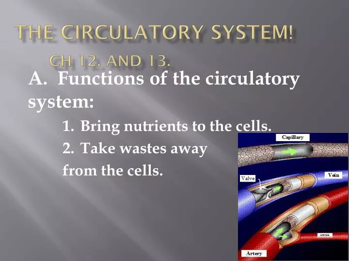
The Circulatory System!Ch 12. and 13. A. Functions of the circulatory system: 1. Bring nutrients to the cells. 2. Take wastes away from the cells.
Five Types of Blood Vessels I. Arteries and arterioles A. Carry blood away from the heart to the tissues. B. Arteries 1. Large, carry blood away from the heart. 2. Thick elastic walls to allow for it to stretch. 3. Surrounded by smooth muscle to control the diameter of the artery. • Circulatory System Overview
Arterioles 1. Arteries branch into arterioles. 2. About 0.2 mm in diameter or smaller. 3. Mostly smooth muscle to allow for more control of the arteriole.
II. Capillaries A. Capillaries connect the arterioles to venules, and exchange material with the tissues.
1. Arterioles branch into small vessels called capillaries. 2. Capillaries are very narrow, microscopic tubes. 3. The walls of these tubes are one cell layer thick. 4. Gases and small molecules like glucose exchange across the walls of the capillaries. 5. In a capillary bed some, many, or most of these sphincter muscles may be closed off so that less or more blood flows to that area, as needed a. e.g. more blood to muscles when they are working. b. e.g. less blood flow to the surface of the skin during hypothermia.
Veins and Venules • Carry blood from the tissues to the heart • Veins • 1. Walls are thinner than arterial walls. • 2. Veins have valves which allow blood to flow only toward the heart when the are open and prevent the backward flow of blood when they are closed. • 3. Act as a blood reservoir. • Venules • 1. Venules join together to form veins • 2. Drain the blood from capillaries and then join to form a vein.
IV. Location of Blood • Veins contain about 75% of the body's blood. • Arteries contain about 20% of the body's blood. • Capillaries contain about 5% of the body’s blood. D. There is close to 100,000 km of blood vessels!
Pulmonary and Systemic Circulation I. Cardiovascular system A. Divided into 2 circuits: 1. PULMONARY CIRCUIT • SYSTEMIC CIRCUIT Overview of P+S Systems
Pulmonary Circuit • A. Path of blood from the heart to/from the lungs. • B. Powered by the right ventricle of the heart. • C. Deoxygenated blood from all tissues collects in the right atrium, is pumped to the right ventricle, then is sent to the pulmonary trunk, which divides into pulmonary arteries, which divide up into the arterioles of the lungs. • D. These arterioles take blood to the pulmonary capillaries, where CO2 and O2 are exchanged. • E. The oxygenated blood then enters pulmonary venules, then the pulmonary veins, and finally back to the left atrium.
III. The Systemic Circuit • A. Includes all blood vessels except those in the pulmonary circuit. • B. Blood is pumped to the tissues and organs by the left ventricle of the heart. • C. From the tissues, blood collects in the right atrium via the superior (anterior) vena cava which drains the head and upper body and the inferior (posterior) vena cava which drains the lower body • D. Blood is then pumped to the lungs through the pulmonary circuit
IV. Oxygenated and Deoxygenated blood • A. In the pulmonary system • 1. Arteries carry deoxygenated blood. • Veins carry oxygenated blood. • B. In the systemic system • 1. Arteries carry oxygenated blood. • 2. Veins carry deoxygenated blood.
Significant Vessels • I. Pulmonary Circuit
Path of a blood cell 1. You should also be able to describe the flow of blood around the body through any major organ!
2. Path of blood to kidneys • Left ventricle to aorta torenal artery to renal arterioles to capillaries to venulesto renal vein toinferior venae cava toright atrium
Path of blood to the intestines a. Left ventricle to aorta to mesenteric artery to capillaries in the villi tovenulesto hepatic portal vein to liver hepatic vein to inferior venae cava to right atrium
Adult and Fetal Circulation I. Fetal Heart Heart develops in 3rd and 4th weeks in uterus. At end of 8 weeks, the embryo’s organ systems, including heart, are functioning. During fourth month, fetal heartbeat is loud enough to be heard with stethoscop Image: Ultrasound showing 4 chamber heart Video: 12 week ultrasound – you can see beating heart
Differences Between Fetal and Adult Circulation • A. Differences • Fetal lungs are NOT used to provide oxygen since it cannot breathe air inside the womb because is immersed in amniotic fluid • 2. Fetus must get all its nutrients from mom, as well as let her take care of its wastes. Click for Animation
Four Features Unique in the Fetus • 1. OVAL OPENING (foramen ovale) • Opening between the right and left atria, covered by a flap that acts like a valve. • Some of the blood from the right atrium is therefore pumped through this flap and into the left atrium, bypassing the pulmonary circuit. • c. If the oval opening doesn’t close after birth, it can cause mixing of blood and “blue babies”. Correct with open heart surgery.
2. ARTERIAL DUCT (ductusarteriosus) • Connects pulmonary artery and aorta. • Much of the blood being pumped out of the heart to the lungs will be directed away from the lungs and into the aorta. • Like the oval opening, the arterial duct’s function is to bypass the pulmonary circuit.
UMBILICAL ARTERIES AND VEINS • Vessels that travel to and from PLACENTA • Placenta is a membrane shared by the mother and baby across which gases, nutrients, and wastes are exchanged • Artery travels toward placenta with waste • c. The umbilical arteries are grafted to the iliac arteries. • d. Vein travels from placenta to fetus with blood rich in O2 and nutrients
4. VENOUS DUCT (ductusvenosus) • Connects umbilical vein to the vena cava to bring the blood back to the baby’s heart. • It attaches right at the babies liver, but bypasses most of the liver. • c. This is why chemicals ingested by the mother can seriously affect the baby
The path of the blood through the fetus • Begin with blood collecting in Right Atrium • From there, blood can go into Left Atrium through Oval opening plus into Right Ventricle through atrioventricle valve. • Right Ventricle to Pulmonary Artery. Most of blood will go through arterial duct into aorta. • D. Aorta to tissue.
Umbilical arteries lead to placenta, where exchange of gases and nutrients take place. • Umbilical vein carries O2 rich blood. • It enters the venous duct, passes through liver. • H. Venous duct joins with inferior vena cava (it mixes here with deoxygenated blood) and this mixed blood goes back to the back to heart.
The Lymphatic System • The lymphatic system is another vascular system in your body. • It is separate from your cardiovascular system because it has its own veins and capillaries. • C. It ultimately connects back with the cardiovascular system because the fluid from the lymphatic system eventually gets sent back into the bloodstream.
Lymphatic system takes up excess tissue fluid (fluid that surrounds cells and tissues) from the tissues and moves into the larger lymphatic vessels and through the lymph nodes and eventually enters the blood through the veins in the neck region. Lymph has no pump of its own so its flow depends on pressure from the blood system and the massaging effect of the muscles. It is a one-way system that starts in the tissues and empties into the cardiovascular system.
II. Lymph • Once fluid enters the lymph vessels it is called LYMPH. • Lymph resembles plasma, but is more diluted (about 5% of proteins and 1% of salts) • C. Formed from bits of blood and other body liquids, called interstitial fluid, that collect in the spaces between cells.
Some of the interstitial fluid goes back into the body through the capillary membrane, but most enters the lymphatic capillaries to become lymph. • Along with this interstitial fluid, the lymph also picks up any particles (cell debris, fat globules, etc) that are too big to be absorbed through the capillary membrane. • F. Lymph contains LYMPHOCYTES which are a type of white blood cell.
III. Main Functions of the Lymphatic System • Transport of excess tissue fluid back to cardiovascular system • Absorption of fat from the intestine and transport to blood • C. Fighting infection • 1. Cleansing lymph • 2. Produce lymphocytes (a type of white blood cell) • 3. Some lymphocytes produce antibodies
IV. Components of the Lymphatic System • A. No lymph “arteries” since there is no “pump” in this system • B. Lymph capillaries take up cell fluids • C. Lymph capillaries drain into lymph veins which have valves for one-way flow • D. Lymph veins join to two main trunks • 1. RIGHT LYMPHATIC DUCT • a. Drains the upper right portion of the body and empties into the right subclavian vein • 2. THORACIC DUCT • a. Drains the rest of the body and drains into the left subclavian vein
V. Other Parts of the Lymphatic System • A. Lacteal • 1. Blind ends of lymph vessels in villi of the small intestine. • Products of fat digestion enter here. • B. Lymph Nodes • 1. Small oval or round structures that occur along strategic places on lymph vessels. • 2. They produce and store lymphocytes • 3. These fight infection by producing antibodies which attach to and “flag” or deactivate foreign proteins • 4. Filter lymph of damaged cells, bacteria and spreading cancer cells as well as debris.
C. Spleen • 1. Located behind the stomach. • Contains white blood cells and stores blood. • D. Thymus Gland • 1. Located in the upper thoracic cavity. • 2. Functions in production and maturation of some lymphocytes. • 3. Decreases in size with age.
Capillary – Tissue Fluid Exchange • I. Exchange of Gases • A. Oxygen • 1. 95% is carried by oxyhaemoglobin (HbO2) • a. 200 million hemoglobin molecules per RBC • Each hemoglobin carries four oxygen molecules • 2. 5% dissolved in plasma
B. Carbon dioxide (CO2) • 1. 9% dissolved in plasma • 2. 27% picks up CO2 to form carbaminohemoglobin (HbCO2). • 3. 64% of CO2 is transported as bicarbonate ion (HCO3-) • a. It is formed after CO2 combines with water, forming carbonic acid which then dissociates. • b. Note the following reaction: • CO2 + H2O H2CO3H+ + HCO3- • c. The enzyme CARBONIC ANHYDRASE speeds up this reaction. • d. The H+ released by reaction changes the blood pH. • e. To prevent this H+ is picked up by the globin portion of hemoglobin (to become HHb) so that pH is maintained.
II. Mechanism of Gas Exchange Intro Animation • Due to a pressure differential between blood pressure and osmotic pressure. • Blood pressure is the pressure of blood in blood vessel would tend to push molecules out of the blood.
At arterial side of a capillary bed, bloodpressure is (40 mm Hg) HIGHER thanblood osmotic pressure (25 mm Hg). • 2. Thus plasma constantly “leaks” out through the walls of the capillaries, forming INTERSTITAL FLUID that bathes tissues. • The interstitial fluid contains water, nutrients, hormones, gases, wastes. • Plasma proteins and blood cells are too big so they are left behind in the capillaries.
Oxygen, sugars and amino acids in the fresh plasma diffuse into/taken up by local cells. D. CO2and waste molecules produced in the tissue cells diffuse out of the tissues and into the interstitial fluid.
Osmotic pressure is the opposing force trying to force molecules into the blood. • At the venule side of the capillary beds, blood pressure is now reduced (10 mm Hg) whereas osmotic pressure is about the same (25mm Hg). • Therefore, water, ammonia, and carbon dioxide laden interstitial fluid is now pulled by osmotic pressure back into the blood vessels tend to enter the bloodstream. • 3. Osmotic pressure is basically constant, but blood pressure varies considerable around a capillary bed. This causes some natural movement of molecules.
K1. Heart Anatomy • Function of the Heart • The pump that circulates the blood throughout the body. • B. A very muscular organ about the size of a fist.