Download
1 / 26
270 likes | 275 Views
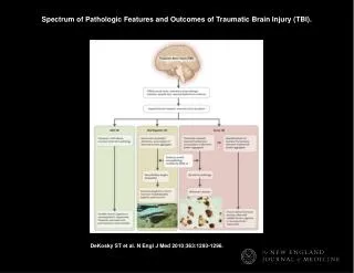
Pathologic Features of Prognostic Significance in Primary Retroperitoneal Liposarcoma. Amanda J. Cannell 1 , Sally M. Burtenshaw 1 , Martin E. Blackstein 2 , Peter Chung 3 , Charles N. Catton 3 , Rebecca A. Gladdy 1 , Carol J. Swallow 1 , Brendan C. Dickson 4.

E N D
Pathologic Features of Prognostic Significance in Primary Retroperitoneal Liposarcoma Amanda J. Cannell1, Sally M. Burtenshaw1, Martin E. Blackstein2, Peter Chung3, Charles N. Catton3, Rebecca A. Gladdy1, Carol J. Swallow1, Brendan C. Dickson4 Departments of Surgery1, Medical Oncology2, Radiation Oncology3, and Pathology and Laboratory Medicine4, Mount Sinai Hospital and Princess Margaret Cancer Centre, University of Toronto; Toronto, ON Canada
INTRODUCTIONRetroperitoneal liposarcoma • Conventional histotypes are well-differentiated (WD) and dedifferentiated (DD) liposarcoma • MDM2 amplification and protein over-expression • Myxoid/ round cell liposarcoma • Pleomorphic liposarcoma • Spindle cell (fibrosarcoma-like)
RATIONALE Predicting behaviour of primary retroperitoneal liposarcoma (RP LPS) is challenging • Conventional indicators not applicable (e.g., TNM) • Local recurrence is predominant clinical challenge • Lack of consensus on prognostic indicators • Clinical and surgical attributes inconsistent • Pathologic • FNCLCC grade • Low-grade (LG-) versus high-grade (HG-) dedifferentiation
RATIONALEBackground - FNCLCC grading system DIFFERENTIATION MITOTIC COUNT NECROSIS
RATIONALEBackground - Differentiation Well-differentiated • Lipoma-like • Sclerosing • Inflammatory Dedifferentiated • Low-grade (LG-DD) • High-grade (HG-DD) DIFFERENTIATION Well-differentiated Dedifferentiated High-grade Low-grade
OBJECTIVE Review of institutional experience with RP LPS to assess variables of potential prognostic significance • Pathologic • Fédération Nationale des Centres de Lutte Contre le Cancer (FNCLCC) grading system • Tumour differentiation, mitotic index, necrosis • Histotype • Well-differentiated vs Dedifferentiated • Extent of dedifferentiation • Low-grade vs high-grade DIFFERENTIATION Well-differentiated Dedifferentiated High-grade Low-grade MITOTIC COUNT NECROSIS
OBJECTIVE Review of institutional experience with RP LPS to assess variables of potential prognostic significance • Pathologic • Fédération Nationale des Centres de Lutte Contre le Cancer (FNCLCC) grading system • Tumour differentiation, mitotic index, necrosis • Histotype • Well-differentiated vs Dedifferentiated • Extent of dedifferentiation • Low-grade vs high-grade DIFFERENTIATION Well-differentiated Dedifferentiated High-grade Low-grade MITOTIC COUNT NECROSIS
OBJECTIVE Review of institutional experience with RP LPS to assess variables of potential prognostic significance • Pathologic • Fédération Nationale des Centres de Lutte Contre le Cancer (FNCLCC) grading system • Tumour differentiation, mitotic index, necrosis • Histotype • Well-differentiated vs Dedifferentiated • Extent of dedifferentiation • Low-grade vs high-grade DIFFERENTIATION Well-differentiated Dedifferentiated High-grade Low-grade MITOTIC COUNT NECROSIS
OBJECTIVE Review of institutional experience with RP LPS to assess variables of potential prognostic significance • Pathologic • Fédération Nationale des Centres de Lutte Contre le Cancer (FNCLCC) grading system • Tumour differentiation, mitotic index, necrosis • Histotype • Well-differentiated vs Dedifferentiated • Extent of dedifferentiation • Low-grade vs high-grade DIFFERENTIATION Well-differentiated Dedifferentiated High-grade Low-grade MITOTIC COUNT NECROSIS
METHODS • Cases identified from prospective database • Jan 1996 to Dec 2013 • Pathology review of all RP sarcomas (pre-treatment biopsy and resections) • College of American Pathologists: synoptic criteria, incl: • FNCLCC Grading • Extent of dedifferentiation, based on WHO criteria • Statistical analysis by SPSS • Chi-squared and Kaplan Meier curves (log-rank)
STUDY POPULATION • Inclusion Criteria • Primary RP LPS treated with curative intent • Unequivocal diagnosis, based on: • Histomorphology • IHC for MDM2 • FISH for MDM2 • Exclusion • Other diagnosis (e.g., lipoma, leiomyosarcoma)
RESULTSSurvival analysis Disease Specific Survival Local Recurrence n=104 % Recurrence % Survival n=104 5 yr: 84% 5 yr: 26% Follow Up Time (Months) Follow Up Time (Months) Median Follow Up Time: 50 Months (5 – 218)
RESULTSHistotype (WD vs DD) Disease Specific Survival Local Recurrence n=30 n=74 % Recurrence n=74 % Survival n=30 p<0.01 p=0.01 Follow Up Time (Months) Follow Up Time (Months)
RESULTSFNCLCC Grade (1, 2, 3) Disease Specific Survival Local Recurrence n=31 n=68 n=5 n=68 % Survival % Recurrence n=5 n=31 p=0.03 p=0.01 Follow Up Time (Months) Follow Up Time (Months)
RESULTSExtent of Differentiation (LG vs HG) Disease Specific Survival Local Recurrence n=56 n=56 n=18 % Recurrence % Survival n=18 p=0.24 p=0.91 Follow Up Time (Months) Follow Up Time (Months)
DISCUSSION • Weaknesses • Small series • Retrospective review • Strengths • Pathology review using modern criteria and diagnostic techniques • Single institution
CONCLUSIONS • Histotype (WD vs DD) is a dominant prognostic indicator DIFFERENTIATION Well-differentiated Dedifferentiated High-grade Low-grade MITOTIC COUNT NECROSIS
CONCLUSIONS • Histotype (WD vs DD) is a dominant prognostic indicator • FNCLCC grade is prognostic • Grade I vs Grade II/III DIFFERENTIATION Well-differentiated Dedifferentiated High-grade Low-grade MITOTIC COUNT NECROSIS
CONCLUSIONS • Histotype (WD vs DD) is a dominant prognostic indicator • FNCLCC grade is prognostic • Grade I vs Grade II/III DIFFERENTIATION High-grade Low-grade Well-differentiated Dedifferentiated High-grade Low-grade MITOTIC COUNT NECROSIS • LG-DD vs HG-DD no prognostic value • Confusing terminology of limited value for RP LPS
Thank you CTOS 2014 Berlin
PATTERNS OF RECURRENCE p = 0.14 p < 0.01