Download
1 / 68
970 likes | 1.98k Views
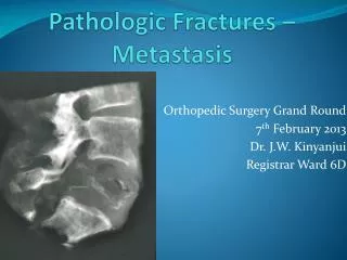
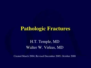
Pathologic Fractures. H.T. Temple, MD Walter W. Virkus, MD Created March 2004; Revised December 2005, October 2008. Pathologic Fractures. Tumors primary secondary (metastatic) (most common) Metabolic osteoporosis (most common) Paget’s disease hyperparathyroidism.

E N D
Pathologic Fractures H.T. Temple, MD Walter W. Virkus, MDCreated March 2004; Revised December 2005, October 2008
Pathologic Fractures • Tumors • primary • secondary (metastatic) (most common) • Metabolic • osteoporosis (most common) • Paget’s disease • hyperparathyroidism
Pathologic Fractures Benign Tumors • Fractures more common in benign tumors (vs malignant tumors) • most asymptomatic prior to fracture • antecedent nocturnal/rest symptoms rare • most common in children • humerus • femur • unicameral bone cyst, NOF, fibrous dysplasia, eosinophilic granuloma
Fractures through benign tumors Unicameral Bone Cyst • Fractures observed more often in males than females • May be active or latent • Almost always solitary • First two decades • Humerus and femur most common sites Fracture through UBC “fallen fragment”sign (arrow)
Unicameral Bone Cyst • Treatment - impending fractures • observation • aspiration and injection methylprednisolone, bone marrow or bone graft • curetting and bone graft (+/-) internal fixation • Treatment - fractures • allow fracture to heal and reassess • ORIF for femoral neck fractures
Fibroxanthoma • Most common benign tumor • Femur, distal tibia, humerus • Multiple in 8% of patients (associated with neurofibromatosis) • Increased risk of pathologic fracture in lesions >50% diameter of bone and >22mm length
Fibroxanthoma • Treatment • observation • curetting and bone graft for impending fractures • immobilization and reassess after healing for patients with fracture
Fibrous Dysplasia • Solitary vs. multifocal (solitary most common) • Femur and humerus • First and second decades • May be associated with café au lait spots and endocrinopathy (Albright’s syndrome)
Fibrous Dysplasia • Treatment • observation • curetting and bone graft (cortical structural allograft) to prevent deformity and fracture (+/-) internal fixation • expect resorption of graft and recurrence • pharmacologic—bisphosphonates
Pathologic Fracturesthrough Primary Malignant Tumors • Relatively rare (often unsuspected) • May occur prior to or during treatment • May occur later in patients with radiation osteonecrosis (Ewing’s, lymphoma) • Osteosarcoma, Ewing’s, malignant fibrous histiocytoma, fibrosarcoma
Pathologic FracturesPrimary Malignant Tumors • Suspect primary tumor in younger patients with aggressive appearing lesions • poorly defined margins (wide zone of transition, lack of sclerotic rim) • matrix production • periosteal reaction • Patients usually have antecedent pain before fracture, especially night pain
Pathologic FracturesPrimary Malignant Tumors • Pathologic fracture complicates but does not mitigate against limb salvage • Local recurrence is higher • Survival is not compromised • Patients with fractures and underlying suspicious lesions or history should be referred for biopsy
B • Pathologic fracture through MFHarising in antecedent infarct • (H&E 100x) Pleomorphic spindledcells with storiform growth pattern A
Pathologic FracturesPrimary Malignant Tumors • Always biopsy solitary destructive bone lesions even with a history of primary carcinoma • Case:A 62 year-old woman with a history of breast carcinoma presented with a pathologic fracture through a solitary proximal femoral lesion
Post- Pre-op Intermediate grade chondrosarcoma *fixation of primary bone tumors must not be performed until proper evaluation has been performed and the diagnosis has been established in order to prevent potential for spread of tumor.
Pathologic FracturesPrimary Malignant Tumors • Treatment • Immobilization • Traction, ex fix, cast • staging • biopsy • adjuvant treatment (chemotherapy) • resection/amputation
Fractures through non-neoplastic bone disease Metabolic Bone Disease • Osteoporosis • insufficiency fractures • Paget’s disease • early and late stages; most fractures occur in the late stage of disease • Hyperparathyroidism • dissecting osteitis • fractures through Brown tumors
Paget’s Disease • Radiographic appearance • Thickened cortices • Purposeful trabeculae • Mixed sclerosis/lysis • Bowing deformities • Joint arthrosis • Fracture • delayed healing • malignant transformation • Treatment • Osteotomy to correct alignment • Excessive bleeding • Joint arthroplasty vs. ORIF Fracture through Pagetic bone (arrow). Transverse fracture suggests pathologic bone.
Hyperparathyroidism • Adenoma • Polyostotic disease • Mental status changes • Abdominal pain • Nephrolithiasis • Polyostotic disease • mixed radiolucent/radiodense Mixedradiodenseandradiolucentlesions Multiple brown tumors in a patient with primary hyperparathyroidism
Hyperparathyroidism • May be secondary to renal failure • secondary • tertiary • Treatment • parathyroid adenectomy • ORIF for fracture • correct calcium Pathologic fracture through brown tumor (arrow)
Fractures in Patients with Metastatic Disease and Myeloma • Aside from osteoporosis, most common causes of pathologic fracture • Fifth decade and beyond • Appendicular sites: femur and humerus most common • All metastatic tumors are not treated the same
Not All Mets Created Equal • Breast – radiosensitive, chemosensitive • Lung – moderately radiosensitive, chemo sensitivity variable • Prostate – radiosentive, chemosensitive • Thyroid – radiosensitive, chemosensitive • Renal – minimally radiosensitive, variable chemosensitivity
Overall Incidence of Metastases to Bone at Autopsy • 70% Jaffe, 1958 • 12% Clain, 1965 • 32% Johnson, 1970 • 21% Dominok, 1982
Incidence of Metastases at Autopsy by Primary Tumor Site Primary Site% metastasis to Bone Breast 50-85 Lung 30-50 Prostate 50-70 Hodgkin’s 50-70 Kidney 30-50 Thyroid 40 Melanoma 30-40 Bladder 12-25
Incidence of Metastases • 60% of patients with early identified cancer may already have metastases • 10-15% of all patients with primary carcinoma will have radiologic evidence of bone metastases during course of disease
Route of Metastases • Contiguous • Hematogenous • most common Destructive lesions in bone from lung carcinoma (arrows)
Mechanism of Metastases • Release of cells from the primary tumor • Invasion of efferent lymphatic or vascular channels • Dissemination of cells • Endothelial attachment and invasion at distant site • Angiogenesis and tumor growth at distant site Metastatic carcinoma In body pedicle junction
Bone Destruction • Early • most important • osteoclast mediated • (RANK L) • Late • malignant cells may be directly responsible
Metastases of Unknown Origin • 3-4% of all carcinomas have no known primary site • 10-15% of these patients have bone metastases
Diagnostic Strategy for Patients with Unknown Primary % Primary Tumor Identified History and Physical 8% Chest X-Ray 43% Chest CT 15% Abdominal CT 13% Biopsy 8% Rougraff, 1993
Defects • Cortical defects weaken bone especially in torsion • Two types • stress riser - smaller than the diameter of bone • open section defect - larger than the diameter of bone…. causes a 90% reduction in load to failure and demand augmentation and fixation
Impending Pathologic Fracture • 61% of all pathologic fractures occur in the femur • 80% are peritrochanteric • fracture in this area results in significant morbidity • historic data on impending pathologic fracture involves the proximal femur
Impending Pathologic Fracture • Parrish and Murray, 1970 • increasing pain with advancing cortical destruction of lesions involving >50% of the shaft diameter • Beals, 1971 • lesions >2.5 cm are at increased risk to fracture • Murray, 1974 • increased fracture with destruction of > one-third of the cortex, pain after radiotherapy
Impending Pathologic Fracture • Fidler, 1981 % shaft destroyed Incidence Fx (%) 0-25% 0% 25-50% 3.7% 50-75% 61% >75% 79% • Conclusion: Patients with tumors destroying >50% of the diameter of bone require prophylactic internal fixation
Indication for ProphylacticInternalFixation • “Harrington criteria” • >50% of diameter of bone • >2.5 cm • pain after radiation • fracture of the lesser trochanter • Limitations • only for proximal femur • doesn’t account for tumor biology Harrington, K.D.: Clin. Orthop. 192: 222, 1985
Mirels Scoring System Score 1 2 3 Site upper limb lower limb peritrochanteric Pain mild moderate functional Lesion blastic mixed lytic Size <1/3 1/3-2/3 >2/3 Score < 7 – no surgery Score > 7 – prophylactic fixation Mirels, H.: Clin. Orthop. 249: 256, 1989.
Adjuvant Treatment • Radiation • Radiation alone • Complete pain relief in 50% • Partial pain relief in 35% • Radiofrequency ablation • Chemotherapy • Hormone treatment • Bisphosphonates
Adjuvant Treatment • Radiation • Radiation alone • Complete pain relief in 50% • Partial pain relief in 35% • Radiofrequency ablation • Chemotherapy • Hormone treatment • Bisphosphonates
Radiation Therapy • Overall 85% response rate • Median duration of pain relief 12-15 weeks • Tumor necrosis followed by collagen proliferation, woven bone formation, and replacement by lamellar bone • Recalcification by 2-3 months • More than half respond within 1-2 weeks • Various dose and fractionization schedules
Radiation Therapy • Townsend, et al., Journal of Clinical Oncology, 1994 • 64 surgical stabilization procedures, 35 with post-op radiation, 29 with no radiation • Functional use of extremity, avoidance of revision surgery, and survival time increased in radiation group
Radiotherapy Post XRT Prostate CA Pre XRT Prostate CA
Bisphosphonates • “Long-term prevention of skeletal complications of metastatic breast cancer with pamidronate: Protocol 19 Aredia Breast Cancer Study Group” • Hortobagyi, et al. Journal of Clinical Oncology, 1998 • “Zoledronic acid reduces skeletal-related events in patients with osteolytic metastases” • Berenson, et al. Cancer 2001
Treatment Objectives in Metastatic Disease • Decrease pain • Restore function • Maintain/restore mobility • Limit surgical procedures • Minimize hospital time • Early return to function (immediate weightbearing)
Pathologic Fracture Survival • 75% of patients with a pathologic fracture will be alive after one year • the average survival is ~ 21 months
Survival Time • Poor prognostic factors • Presentation with metastatic disease • Short time from initial diagnosis to first met • Visceral mets • Non-small cell lung cancer
Healing of Path Fractures • Healing rate of pathologic fractures • Myeloma- 67% • Renal- 44% • Breast- 37% • Lung- 0%
Fracture Healing • 129 patients • overall rate = 35% • 74% for patients surviving > 6 months • radiotherapy <30 GY did not adversely affect fracture healing Gainor, B.J.: CORR 178: 297, 1983
Pathologic Fracture Treatment • Biopsy especially for solitary lesions • Nails versus plates versus arthroplasty • plates, screws and cement superior for torsional loads • interlocked nails stabilize entire bone • Cement augmentation • Radiation/chemotherapy/bisphosphonates • Aggressive rehabilitation
Indications for Surgical Treatment • Ratio of survival time to surgical recovery time • Ability to ambulate • Ability to use extremity • Capacity to return to full function • Pain not controlled by analgesics • Location of disease – high risk area
Indications for ORIF/IMN • Diaphyseal lesion • Good bone stock • Histology sensitive to chemo/radiation • Impending fractures • Poor prosthetic options