Download
1 / 41
430 likes | 619 Views
The use of flow cytometry in lung diseases. Milton D. Rossman, M.D. Professor of Medicine Hospital University of Pennsylvania. Flow cytometers are made of basic subunits. Workstation Computer Software Data storage Sampling system Flow cell and sensing region Laser/s Optics Electronics.

E N D
The use of flow cytometry in lung diseases Milton D. Rossman, M.D. Professor of Medicine Hospital University of Pennsylvania
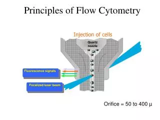
Flow cytometers are made of basic subunits • Workstation • Computer • Software • Data storage • Sampling system • Flow cell and sensing region • Laser/s • Optics • Electronics
Injector Tip Sheath fluid Fluorescence signals Focused laser beam Flow Cell
Laser FALS Sensor Forward Angle Light Scatter
Laser FALS Sensor 90LS Sensor 90 Degree Light Scatter
FALS Sensor Fluorescence Fluorescence detector (PMT3, PMT4 etc.) Fluorescence Detectors Laser
Flow Cytometry Optics PMT 4 PMT Dichroic 3 Filters Flow cell PMT 2 Bandpass Filters PMT 1 Laser
Fluorescently labelled molecules useful in flow cytometry • Monoclonal antibodies • Non-specific protein binding molecules • DNA binding molecules
Disease for which flow cytometry may be useful • Lymphoma • Interstitial lung disseases • Sarcoidosis • Chronic beryllium disease • Hypersensitivity pneumonitis
B-cell tumors represent clonal outgrowth of B-cells at various stages of development
Lymph node Bone marrow HEV Cortex (B-rich follicles) Medulla (T-rich and DCs) Stem cell Acute Leukemia Pro-B Recirculate (efferent lymph) Pre-B Encounter Ag No Ag Peripheral Blood Cell death Immature B-cell B-NHL TH Activation Cell division Follicle with a germinal center = Secondary lymphoid follicle Mature naïve B-cell Marginal zone (Memory B cells) Medulla (differentiate to plasma cells) Multiple Myeloma B-cell malignancies BCR+
B-cell maturation is association with different antigens at each stage Pro-genitor-B Pre Pre-B Plasma-cytoid B Plasma Cell Stem cell Pre-B Pre-B Early-B Mature-B Ia+ CD19+ CD20+ Cd22+ sIg+ Ia+ CD19+ CD20+ Cd22+ sIg+ Ia+ CD19+/- CD20+/- Cd22+/- PCA-1+/- sIg+/- cIg+/- Ia+/- PCA-1+ cIg+ TdT+ TdT+ Ia+ TdT+ Ia+ CD19+ cIg+ TdT+ Ia+ CD19+CD10+ TdT+ Ia+ CD19+CD10+ CD20+
What causes B-NHL?? • Viruses • EBV • Burkitt’s lymphoma • Post-Tx lymphoma • Classic Hodgkin’s disease • Mistakes in gene recombination • Frequently involve Ig heavy and light chain loci • NHL increased incidence in chronic inflammatory conditions (eg. Celiac disease, IBD, Sjögren’s syndrome) • Signature translocations for NHL subtypes
Idiotype Ag binding site Fab Fc BCR - the signature molecule on a mature B cell
Increases Ab affinity for Ag Rescues auto-reactive B-cells Alters the effector function of of Ab Molecular processes modifying the genes encoding the BCR BM/LN Lymphoid organs
How is flow cytometry useful in suspected cases of lymphoma? • Aids diagnosis • Identify clonal B cells (kappa or lambda light chain restricted) • Identify aberrant pattern of antigen expression • Asynchronous • T or myeloid cell marker present on B cells • Monitor the success of therapy • Look for minimal residual disease in BM, PB
Tissue samples that can be used for flow cytometry • Biopsy (lung or mediastinal) • Bronchoalveolar lavage fluid • Fine needle aspiration
Flow cytometry of tissue • Cells are removed from the connective tissue stroma and washed • Stained with a panel of monoclonal antibodies to cell surface proteins that define T vs B cells and their stages of differentiation • Data acquired on the flow cytometer • Multiparameter analysis on a computer to identify the presence or absence of malignant cells
SS vs CD45 gating can be used to detect the lymphoma cells to clearly assess surface light chain and lineage markers B cells: CD19, CD20, CD23, sIg,CD5,CD10 T cells: CD3, CD4, CD8, CD5, CD2, CD16/56 Lymphoma cells may be found in either of these 2 regions CD45 Side scatter
Normal B cells express polyclonal light chains CD19 Kappa Lambda Surface expression of BCR light chain
B- cell lymphoma are light-chain restricted CD19 Kappa Lambda Surface expression of BCR light chain
Malt type, B cell lymphoma identified by BAL • 3 patients with biopsy proven MALT had BAL • Cytological features of lymphoma present in only 2 • Flow cytometry • High percentage of B cells (52, 40, 28%) • Altered Kappa/Lamba ratio in two (38 and 0.04) • Poletti et al, Monaldi Arch Chest Dis. 50:191-194, 1995
Diagnosis of MALT using tranbronchial Bx, FNA, Core needle and wedge excisions • 13 cases diagnosed as MALT • B cell monoclonality established by single light chain in 12/13 • All displayed low S phase consistent with a low grade process • Zaer et al, Modern Path. 11:525-532, 1998
(A-C) B-cell Lymphoma (D-F) Normal polyclonal B cells
Peripheral T-cell lymphoma express a characteristic phenotype
Identification of subpopulations of T cells in BAL fluid • Finding of lymphocytosis may be first indication of sarcoidosis • Increased CD4/CD8 ratio (> 4.0) may be relatively specific for sarcoidosis • Eur Respir J. 10;2722, 1997 • Eur Respir J. 10;2716, 1997 • Eur Respir J. 10;2699, 1997 • Need to eliminate CBD and Mycobacterial diseases
Chronic Beryllium Disease • Chronic granulomatosis involving lungs and occasionally the skin. • Only occurs in individuals with beryllium exposure.
CBD can lead to end stage lung disease • Cyst formation • Distortion of airways • Large calcified nodes
Typical trans-bronchial biopsy in CBD • Epitheliod granuloma • Multinucleated Giant cell • Schumann body • Lymphocytic infiltrates
Epidemiological evidence 1. Low attack rate ~ 5% 2. No clear cut dose-reponse relationship 3. Granulomatous reaction 4. Delayed skin reactivity Immunological evidence 1. Increased CD4+ T cells in the lung 2. Positive Be LPT in all individuals 3. Increased Be specific cells in the lungs CBD is due to a hypersensitivity to beryllium.
Flow cytometry for cellular proliferation • CFSE – binds to protein and is divided equally when the cell divides • Can combine with surface labeling to identify the phenotype of the dividing cell
Comparison of 3H-thy with CSFE flow cytometry • 9 Normal Controls • 24 Individual with prior evidence of beryllium sensitivity • 3H-thymidine uptake by stimulation index • CFSE ratio of divided to undivided cells • CD3, CD4 subpopulations
After 7 days of incubation, proliferation could be easily detected
Potential uses of CSFE for measuring cellular proliferation • Chronic beryllium disease • Other hypersensitivity pneumonitis • Metal induced allergic responses • Status of cell mediated immunity in HIV and in patients with immunodeficiencies
Current uses of flow cytometry in lung disorders • Diagnosis of lymphoma • Identification of lymphocyte subpopulations
Future uses of flow cytometry in lung disorders • Identification of specific cell proliferation for the specific diagnosis of hypersensitivity pneumonitis • Identification of lung malignancy