Download
1 / 15
160 likes | 363 Views
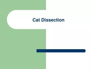
Anatomy: Cat Dissection Part II. Dissection of the superior extremity. Table of Contents. Slide 14 – Pectoantebrachialis Slide 15 – Pectoralis Slide 16 – Combined View Slide 17 – Xiphi Humeralis Slide 18 – Epitrochlearis Slide 19 – Rhomboideus Slide 20 – Serratus Anterior

E N D
Anatomy: Cat Dissection Part II Dissection of the superior extremity
Table of Contents Slide 14 – Pectoantebrachialis Slide 15 – Pectoralis Slide 16 – Combined View Slide 17 – Xiphi Humeralis Slide 18 – Epitrochlearis Slide 19 – Rhomboideus Slide 20 – Serratus Anterior Slide 21 – Serratus Dorsalis Slide 22 – Levator Scapulae Slide 4 – External Jugular Vein Slide 5 – Sternomastoid Slide 6 – Cleidomastoid Slide 7 – Submandibular Salivary Gland Slide 8 - Parotid Salivary Gland Slide 9 – Clavicular Trapezius Slide 10 – Acromial Trapezius Slide 11 – Spino Trapezius Slide 12- Acromial Aponeurosis Slide 13 – Latissimus Dorsi Slide 23 – Transverse Cervical Artery Slide 24 – Transverse Cervical Vein Slide 25 – Clavicular Deltoid Slide 26 – Acromical Deltoid Slide 27 – Spino Deltoid Slide 28 – Supraspinatus Slide 29 – Infraspinatus
Table of Contents (Con.) Slide 30 – Teres Minor Slide 31 - Teres Major Slide 32 – Subscapularis Slide 33 - Coracobrachialis Slide 34 – Triceps Brachii long head Slide 35 – Triceps Brachii lateral head Slide 36 - Triceps Brachii medial head Slide 37 – Anconeus Slide 38 – Biceps Brachii Slide 39 – Brachialis Slide 40 – Brachioradialis Slide 41 – Extensor Digitorum Communis Slide 42 – Extensor Digitorum Lateralis Slide 43 - Extensor Carpi Radialis Slide 44 – Supinator Slide 45 - Extensor Pollicis Brevis Slide 46 – Extensor Carpi Ulnaris Slide 47 – Extensor Indicis Slide 48 – Pronator Teres Slide 49 – Palmaris Longus Slide 50 – Flexor Carpi Radialis Slide 51 - Flexor Carpi Ulnaris Slide 52 – Flexor Digitorum Profundus Slide 53 – Flexor Digitorum Sublimis Slide 54 – Pronator Quadratus
External Jugular Vein Purpose: Carry blood from head Superior inferior
Sternomastoid O: Sternum I: Skull P: Turns head laterally
Cleidomastoid O: Sternum I: Skull P: Turns head laterally
Submandibular gland This gland produces saliva.
Parotid gland This gland also produces saliva.
ClavicularTrapezius O: Cervical and Thoracic Vertebrae I: Scapula and Clavicle P: Elevate Shoulder
Acromial Trapezius O: Cervical and Thoracic Vertebrae I: Scapula and Clavicle P: Retracts Scapula
Spino Trapezius O: Cervical and Thoracic Vertebrae I: Scapula and Clavicle P: Depress Shoulder
Acromial Aponeurosis Acromial Aponeurosis is visible at the top of the photo(8). It originates at the junction of the Right & Left acromio-trapezius and its purpose is to allow for flexibility.
Latissimus Dorsi O: Lumbodorsal aponeurosis I: Humerus P: Adducts humerus
Pectorantebrachialis O: Manubrium I: Forearm P: Adducts Forearm
Pectoralis O: Sternum, ribs I: Humerus, Scapula P: Adducts arm