Download
1 / 26
260 likes | 266 Views
Chapter 46: Animal Reproduction. What are the 2 ways that animals reproduce? Asexual – mitosis – same genes as parent Fission – separation of a parent into 2 or individuals of the same size Budding – individuals split as an outgrowth from an existing one Some cnidarians

E N D
Chapter 46: Animal Reproduction • What are the 2 ways that animals reproduce? • Asexual – mitosis – same genes as parent • Fission – separation of a parent into 2 or individuals of the same size • Budding – individuals split as an outgrowth from an existing one • Some cnidarians • Fragmentation – breaking of the body into several pieces followed by • regeneration • Sponges, cnidarians, tunicates • Sexual – fusion of haploid gametes • Motile sperm swims to non-motile egg • Increases genetic variability • What are the 2 types of sexual reproduction? • External – eggs shed by female & fertilized by male in wet environment • Courtship behaviors involved • Pheromones used • Internal - sperm deposited in or near female reproductive tract • & fertilized within female • Fewer zygotes but more parental care • Embryo develops in reproductive tract
Eggs Figure 46.5 External fertilization
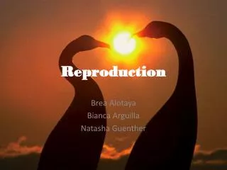
Chapter 46: Animal Reproduction • What are the 2 ways that animals reproduce? • What are the 2 types of sexual reproduction? • External – eggs shed by female & fertilized by male in wet environment • Courtship behaviors involved • Pheromones used • Internal - sperm deposited in or near female reproductive tract • & fertilized within female • Fewer zygotes but more parental care • Embryo develops in reproductive tract • What is parthenogenesis? • Process in which an egg develops without fertilization • Produces haploid adults that produce eggs without meiosis • Daphnia (water flea), bees (male drones), wasps, ants
(a) Both lizards in this photograph are C.uniparensfemales. The one on top is playing the role of a male. Every two or three weeks during the breeding season, individuals switch sex roles. Ovarysize Ovulation Ovulation Progesterone Estrogen Hormones (b) The sexual behavior of C. uniparens is correlated with the cycle of ovulation mediated by sex hormones. As blood levels of estrogen rise, the ovaries grow, and the lizard behaves like a female. After ovulation, the estrogen level drops abruptly, and the progesterone level rises; these hormone levels correlate with male behavior. Time Behavior Female-like Male-like Female-like Male-like Figure 46.3 Sexual behavior in parthenogenetic lizards
Chapter 46: Animal Reproduction • What are the 2 ways that animals reproduce? • What are the 2 types of sexual reproduction? • What is parthenogenesis? • What does the male reproductive tract look like?
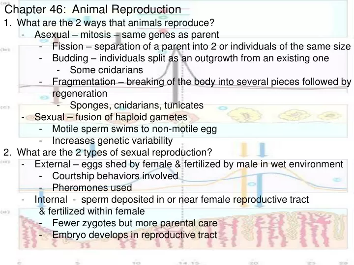
(Urinary bladder) Seminal vesicle (Rectum) (Pubic bone) Erectile tissue of penis Vas deferens Ejaculatory duct Prostate gland Urethra Bulbourethral gland Glans penis Vas deferens Epididymis Testis Prepuce Scrotum Figure 46.10 Reproductive anatomy of the human male External – scrotum & penis Internal – gonads produce sperm & hormones - testes – packed with highly coiled seminiferous tubules - accessory glands that help sperm movement - seminal vesicles - bulbourethral glands - prostate gland
(Urinarybladder) Seminal vesicle (behind bladder) Prostate gland Bulbourethral gland Urethra Erectile tissue of penis Scrotum Vas deferens Epididymis Glans penis Testis 5. Where do the sperm travel during ejaculation? Propelled from epididymis vas deferens ejaculatory duct 6. How many sperm are released during ejaculation? 2 – 5 mL of semen per ejaculation x 50 – 130 million sperm per mL 100 – 650 million sperm per ejaculation
What happens after ejaculation? • Prostaglandins in semen thin mucus as the opening of the uterus & stimulate uterine contractions which help semen move up uterus • Semen (slightly alkaline) neutralizes vagina (slightly acidic) • Protects sperm & increases motility • Initially sperm are coagulated & become liquified so sperm can swim • How does viagra work? • Promotes the action of nitric oxide (NO) which enhances relaxation of smooth muscle in blood vessels of penis so blood can enter erectile tissue 9. What does the female reproductive anatomy look like?
Uterus (Urinary bladder) Oviduct (Pubic bone) Ovary (Rectum) Cervix Vagina Urethra Shaft Prepuce Clitoris Bartholin’s gland Glans Labia minora Vaginal opening Labia majora Figure 46.9 Reproductive anatomy of the human female External – clitoris & 2 sets of labia which surround the clitoris & vaginal opening Internal – pair of gonads (ovaries) with ducts & chambers for gametes & fetus
Oviduct Ovaries Follicles Uterine wall Uterus Cervix Endometrium Corpus luteum Vagina • Ovaries – contain many follicles • Follicle – all formed before birth • egg cell surrounded by follicle cells which nourish & protect the egg • produce estrogens • Endometrium – inner lining of uterus • After ovulation, “egg” falls down the oviduct into uterus
What happens after ejaculation? • Prostaglandins in semen thin mucus as the opening of the uterus & stimulate uterine contractions which help semen move up uterus • Semen (slightly alkaline) neutralizes vagina (slightly acidic) • Protects sperm & increases motility • Initially sperm are coagulated & become liquified so sperm can swim • How does viagra work? • Promotes the action of nitric oxide (NO) which enhances relaxation of smooth muscle in blood vessels of penis so blood can enter erectile tissue • What does the female reproductive anatomy look like? • How are “eggs” made? - Oogenesis
Ovary Primary germ cell in embryo Differentiation Oogonium in ovary Oogonium 2n Mitotic division Primary oocyte within follicle Primary oocyte, arrested in prophase of meiosis I (present at birth) 2n Growing follicle Completion of meiosis I and onset of meiosis II n First polar body Secondary oocyte, arrested at meta- phase of meiosis II n Ovulation Mature follicle Entry of sperm triggers completion of meiosis II n n Ruptured follicle Ovum Ovulated secondary oocyte Corpus luteum Degenerating corpus luteum Figure 46.11 Human Oogenesis Follicles grow in response to FSH completing meiosis I & stopping at metaphase II Smaller polar body is discarded Secondary oocyte is ovulated…NOT AN EGG Meiosis is completed IF 2° oocyte is fertilized After ovulation, remaining tissue reorganizes to form the corpus luteum Second polar body
What happens after ejaculation? • Prostaglandins in semen thin mucus as the opening of the uterus & stimulate uterine contractions which help semen move up uterus • Semen (slightly alkaline) neutralizes vagina (slightly acidic) • Protects sperm & increases motility • Initially sperm are coagulated & become liquified so sperm can swim • How does viagra work? • Promotes the action of nitric oxide (NO) which enhances relaxation of smooth muscle in blood vessels of penis so blood can enter erectile tissue • What does the female reproductive anatomy look like? • How are “eggs” made? • Oogenesis 11. How are sperm made? • Spermatogenesis
Epididymis Seminiferous tubule Testis Cross section of seminiferous tubule 2n Spermatogonium Sertoli cell nucleus Mitotic division, producing large numbers of spermatogonia Differentiation and Onset of meiosis I Primary spermatocyte (in prophase of meiosis I) 2n Meiosis I completed n n Secondary spermatocyte Meiosis II Lumen of Seminiferous tubule Spermatids (at two stages of differentiation) Early spermatids n n n n Differentiation (Sertoli cells provide nutrients) Sperm cells n n Neck n n Head Midpiece Tail Plasma membrane Mitochondria Nucleus Acrosome Figure 46.12 Human Spermatogenesis • - Seminiferous tubules – produce sperm • Leydig cells – secrete testosterone & • other androgens • Sertoli cells – provide nutrition for • spermatids so they can mature • Takes 65 – 75 days to make sperm • 20 days to travel epididymis where • they mature & are stored
What happens after ejaculation? • Prostaglandins in semen thin mucus as the opening of the uterus & stimulate uterine contractions which help semen move up uterus • Semen (slightly alkaline) neutralizes vagina (slightly acidic) • Protects sperm & increases motility • Initially sperm are coagulated & become liquified so sperm can swim • How does viagra work? • Promotes the action of nitric oxide (NO) which enhances relaxation of smooth muscle in blood vessels of penis so blood can enter erectile tissue • What does the female reproductive anatomy look like? • How are “eggs” made? • Oogenesis 11. How are sperm made? • Spermatogenesis • What are the differences between spermatogenesis & oogenesis? • Unequal cytokinesis in oogenesis produces 1 haploid ovum (& 2 polar bodies) & not 4 haploid sperm • Spermatogenesis mitosis occurs throughout life but ovary has all its follicles at birth • Oogenesis has long resting periods but spermatogenesis is uninterrupted & continuous 13. How do hormones control the menstrual cycle?
Control by hypothalamus (a) Inhibited by combination of estrogen and progesterone Hypothalamus Stimulated by high levels of estrogen GnRH 1 Anterior pituitary Inhibited by low levels of estrogen LH FSH 2 (b) Pituitary gonadotropins in blood 6 LH FSH FSH and LH stimulate follicle to grow LH surge triggers ovulation 3 (c) Ovarian cycle 7 8 Corpus luteum Degenerating corpus luteum Growing follicle Mature follicle Ovulation Luteal phase Follicular phase Progesterone and estrogen secreted by corpus luteum Estrogen secreted by growing follicle in increasing amounts 4 Peak causes LH surge (d) Ovarian hormones in blood 5 10 Progesterone Estrogen 9 Progesterone and estro- gen promote thickening of endometrium Estrogen level very low (e) Uterine (menstrual) cycle • Endometrium Secretory phase Menstrual flow phase Proliferative phase 25 28 5 14 15 0 20 10 Days Figure 46.13 The reproductive cycle of the human female GnRH – Gonadotropin Releasing Hormone - hypothalamus - stimulates release of FSH & LH by anterior pituitary FSH - Follicle Stimulating Hormone - follicle grows & expresses LH receptors LH - Lutenizing Hormone - triggers ovulation (1 day after LH peak) - stimulates formation of corpus luteum Estrogen – produced by growing follicle - stimulates GnRH release which triggers more LH & FSH - estrogen peak causes LH peak Progesterone – from corpus luteum - maintains thick endometrium in preparation for embryo implant If pregnancy does not occur the endometrium is shed….menstruation. Menopause – cessation of ovulation & menstru- ation because ovaries lose responsive- ness to LH & FSH & estrogen decreases
What happens after ejaculation? • How does viagra work? • What does the female reproductive anatomy look like? • How are “eggs” made? 11. How are sperm made? • What are the differences between spermatogenesis & oogenesis? • How do hormones control the menstrual cycle? • How do hormones control the male reproductive system? - GnRH, FSH, & LH
Stimuli from other areas in the brain Hypothalamus GnRH from the hypothalamus reg- ulates FSH and LH release from the anterior pituitary. Anterior pituitary Negative feedback FSH acts on the Sertoli cells of the seminiferous tubules, promoting spermatogenesis. LH stimulates the Leydig cells to make testosterone, which in turn stimulates sperm production. Leydig cells make testosterone Primary and secondary sex characteristics Sertoli cells Spermatogenesis Testis Figure 46.14 Hormonal control of the testes
What happens after ejaculation? • How does viagra work? • What does the female reproductive anatomy look like? • How are “eggs” made? 11. How are sperm made? • What are the differences between spermatogenesis & oogenesis? • How do hormones control the menstrual cycle? • How do hormones control the male reproductive system? - GnRH, FSH, & LH • What is detected in pregnancy tests? • HCG – human chorionic gonadotropin • Mimics LH which keeps corpus luteum secreting estrogen & progesterone • Endometrium stays thick • What happens after fertilization?
Cleavage (cell division) begins in the oviduct as the embryo is moved toward the uterus by peristalsis and the movements of cilia. Cleavage continues. By the time the embryo reaches the uterus, it is a ball of cells. It floats in the uterus for several days, nourished by endometrial secretions. It becomes a blastocyst. 3 4 Ovary Fertilization occurs. A sperm enters the oocyte; meiosis of the oocyte finishes; and the nuclei of the ovum and sperm fuse, producing a zygote. 2 The blastocyst implants in the endometrium about 7 days after conception. 5 Uterus Ovulation releases a secondary oocyte, which enters the oviduct. 1 Endometrium (a) From ovulation to implantation Endometrium Inner cell mass Cavity Trophoblast Blastocyst (b) Implantation of blastocyst Fig 46.15 Formation of the zygote and early post-fertilization events Inner cell mass – develops into embryo & extra-embryonic membranes Trophoblast – outer layer of blastocyst that grows out & mingles with endometrium & helps form the placenta Placenta – disk shaped organ containing embryonic & maternal blood vessels
Maternal veins Maternal arteries Placenta Maternal portion of placenta Umbilical cord Chorionic villus containing fetal capillaries Fetal portion of placenta (chorion) Maternal blood pools Uterus Umbilical arteries Fetal arteriole Umbilical vein Fetal venule Umbilical cord Figure 46.16 Placental circulation As placenta forms, HCG levels decline & placenta secretes its own progesterone Fetal nutrients gained from maternal blood pools….no mixing of blood.
What happens after ejaculation? • How does viagra work? • What does the female reproductive anatomy look like? • How are “eggs” made? 11. How are sperm made? • What are the differences between spermatogenesis & oogenesis? • How do hormones control the menstrual cycle? • How do hormones control the male reproductive system? - GnRH, FSH, & LH • What is detected in pregnancy tests? • HCG – human chorionic gonadotropin • Mimics LH which keeps corpus luteum secreting estrogen & progesterone • Endometrium stays thick • What happens after fertilization? • How do hormones regulate parturition, aka child birth?
Estrogen Oxytocin from fetus and mother's posterior pituitary from ovaries Positive feedback Induces oxytocin receptors on uterus Stimulates uterus to contract Stimulates placenta to make Prostaglandins Stimulate more contractions of uterus Figure 46.18 A model for the induction of labor
Placenta Umbilical cord Uterus Cervix 1 Dilation of the cervix 2 Expulsion: delivery of the infant Uterus Placenta (detaching) Umbilical cord 3 Delivery of the placenta Figure 46.19 The three stages of labor
What happens after ejaculation? • How does viagra work? • What does the female reproductive anatomy look like? • How are “eggs” made? 11. How are sperm made? • What are the differences between spermatogenesis & oogenesis? • How do hormones control the menstrual cycle? • How do hormones control the male reproductive system? - GnRH, FSH, & LH • What is detected in pregnancy tests? • HCG – human chorionic gonadotropin • Mimics LH which keeps corpus luteum secreting estrogen & progesterone • Endometrium stays thick • What happens after fertilization? • How do hormones regulate parturition, aka child birth? • How does contraception work?
Female Male Event Event Method Method Production of viable sperm Production of viable oocytes Vasectomy Combination birth control pill (or injection, patch, or vaginal ring) Sperm transport down male duct system Ovulation Abstinence Abstinence Condom Coitus interruptus (very high failure rate) Sperm deposited in vagina Capture of the oocyte by the oviduct Tubal ligation Spermicides; diaphragm; cervical cap; progestin alone (minipill, implant, or injection) Transport of oocyte in oviduct Sperm movement through female reproductive tract Meeting of sperm and oocyte in oviduct Morning-after pill (MAP) Union of sperm and egg Progestin alone Implantation of blastocyst in properly prepared endometrium Birth Figure 46.20 Mechanisms of some contraceptive methods Birth control pills – estrogen & progesterone combo - prevents ovulation by decreasing the release of GnRH which inhibits FSH & LH Morning after pill – higher doses of estrogen & progesterone - prevents fertilization or implantation