Download
1 / 23
240 likes | 491 Views
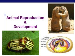
Animal Reproduction & Development Ch . 46-47. Asexual Reproduction vs. Sexual Reproduction Sexual Reproduction : offspring form by fusion of haploid gametes (egg & sperm cells) to make a diploid zygote.

E N D
Animal Reproduction & Development Ch. 46-47
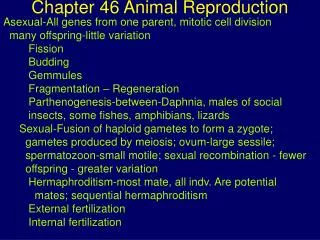
Asexual Reproduction vs. Sexual Reproduction • Sexual Reproduction:offspring form by fusion of haploid gametes (egg & sperm cells) to make a diploid zygote. • Asexual Reproduction: all genes come from one parent; no fusion of egg and sperm.
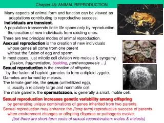
Advantages to asexual reproduction • Animals in isolation can reproduce without finding mates • Creates large numbers of offspring in a short time (can colonize habitats rapidly) • Most advantageous in stable, favorable environments because it perpetuates successful genotypes exactly. • Hermaphroditism: each individual has both male and female reproductive systems--can have dual fertilization when 2 organisms mate (both will release sperm and eggs; results in twice the offspring) • ex. earthworms, parasites such as tapeworms--organisms that seldom encounter the opposite sex.
Costs and Benefits of Sexual Reproduction Costs--Energy use for: • producing, nurturing, and delivering gametes • specialized reproductive systems • investments in courtship of a mate • reproductive timing controls (think spring zoo babies!) • locating and recognizing mates--through pheromones, visual signals such as colors/patterns on fur or feathers, territorial defense rituals, etc. • Ensuring survival of offspring, depending on number of offspring produced--parental care levels • Nourishment of developing organism Benefits of Sexual Reproduction • Biggest benefit: diversity among organisms--variations in the traits of offspring-- to provide evolutionary adaptations and success.
Meiosis in mammals • Spermatogenesis: produces 4 haploid, motile sperm cells from diploid parent cells called spermatogonia. • Oogenesis: developments of 1 mature egg cell, called an ovum, and 3 polar bodies. Oogonia are the diploid parent cells that develop into ova. • Oogonia start to multiply early in female embryonic development, but stop at prophase I in meiosis I and are called primary oocytes (still diploid). • At puberty, primary oocytes are stimulated by FSH to grow in a follicle (in ovary)_and continue meiosis – makes a secondary oocyte, which can be fertilized by a sperm cell once released.
Spermatogenesis v. Oogenesis • 4 sperm cells produced v. a single egg per meiotic division. • Spermatogenesis continues from puberty throughout life in males; oogenesis ends at menopause. • Sperm are produced continuously after puberty while eggs are produced only once per month after puberty.
Ovarian Cycle • Follicular Phase – follicle with immature egg cell develops, secreting estrogen • Ovulation – unfertilized egg cell is released • Luteal Phase – remaining follicle, now the corpus luteum breaks down, secreting estrogen and progesterone
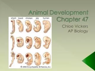
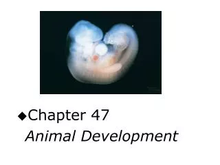
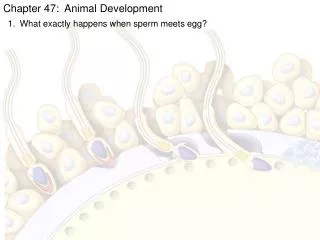
Ch. 47: Animal Developmental Stages 1. Gamete formation: oogenesis and spermatogenesis 2. Fertilization: egg and sperm fuse, nuclei fuse to form a zygote 3. Cleavage: mitotic cell divisions that divide the volume of egg cytoplasm into smaller daughter cells, called blastomeres. (No growth yet, just increasing the number of cells) Blastocoel: Fluid-filled central cavity
In mammals, the blastula is called a blastocyst. -Inner cell mass is a group of cells that develop into the embryo. The cells are the embryonic stem cells. -Trophoblastis outer layer of blastocyst, which forms the fetal part of placenta.
Developmental Stages cont'd. 4. Gastrulation: major cellular reorganization. The new cells are arranged into a gastrula with 2 or 3 primary tissues (germ layers) which will give rise to all tissues and organs of the adult organism.
Developmental Stages cont'd. 5. Organ formation (organogenesis): the primary tissue layers form subpopulations of cells, which become specialized organs and tissues in exact spatial patterns and at specific times. 6. Growth and tissue specialization: tissues and organs grow to final size, shape, proportions, and assume their specific functions--this stage extends into adulthood.
Morphogenesis: a program of orderly changes in an embryo's size, shape, and proportions. Cells divide, grow, migrate, and change size. Tissues expand and fold; some cells die in controlled ways (apoptosis!) at certain locations... think fingers vs. webbed hands. neural tube
The Division of the Gray Crescent • Control group – normal division of cells, dividing the gray crescent evenly between the new cells = normal offspring • Experimental group – induction of cell division (using a thread) so that the gray crescent was all on one side and only one blastomere received the gray crescent = one belly piece and one normal offspring • Conclusion – the gray crescent contains cytoplasmic determinants that influence normal development of offspring from the first cell division
Cell Differentiation: when a cell selectively activates genes and makes proteins not found in other cell types. • -Embryonic cells are totipotent, meaning they are capable of developing into ALL the different cells types in a species. • -Every cell of an embryo has the same DNA, but each cell activates particular genes and not others. • Patterns of development are due to cytoplasmic determinants (chemical signals such as mRNAs) and inductive cell signals. • Embryonic induction: developmental fates of embryonic cell lineages will change when exposed to gene products from neighboring tissues. • Thus, embryonic cells receive signals and begin to form specialized tissues and organs in ordered, spatial patterns.