Download
1 / 1
10 likes | 144 Views
Nano-Composites for Load-Bearing Applications C. Freeman 1 , S. Grubb 2 , N. Cummins 3 , C. Reidy 3 , D. Curran 3 , S. Hampshire 3 , I. Brook 2 and M. Towler 4 1 Charles Clifford Dental Hospital, Sheffield, UK, 2 University of Sheffield, UK, 3 University of Limerick, Ireland,

E N D
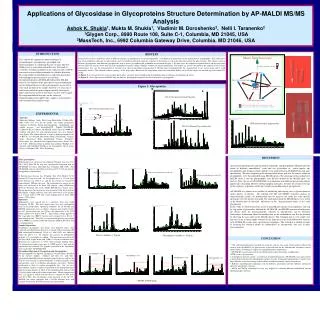
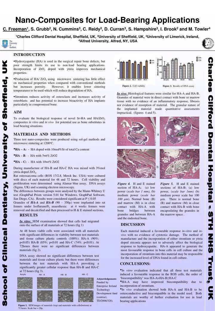
Nano-Composites for Load-Bearing Applications C. Freeman1, S. Grubb2, N. Cummins3, C. Reidy3, D. Curran3, S. Hampshire3, I. Brook2 and M. Towler4 1Charles Clifford Dental Hospital, Sheffield, UK, 2University of Sheffield, UK, 3University of Limerick, Ireland, 4Alfred University, Alfred, NY, USA • INTRODUCTION • Hydroxyapatite (HA) is used in the surgical repair bone defects, but poor strength limits its use to non-load bearing applications. Incorporation of ZrO2 doped with yttria improves mechanical properties. • Production of HA/ ZrO2 using microwave sintering has little effect on mechanical properties when compared with conventional methods but increases porosity. However, it enables lower sintering temperatures to be used which will reduce degradation of HA. • Strontium reduces activity of osteoclasts and increases activity of osteoblasts and has potential to increase bioactivity of HA implants particularly in compromised bone. • AIM • To evaluate the biological response of novel Sr-HA and HA/ZrO2 composites in vitro and in vivo for potential use as bone substitutes in load bearing situations. • MATERIALS AND METHODS • Three test nano-composites were produced using sol-gel methods and microwave sintering at 1200oC. • HA – A: - HA doped with 10mol%Sr of total Ca content • HA – B: - HA with 5wt% ZrO2 • HA – C: - HA with 10wt% ZrO2 • During manufacture of HA-B and HA-C HA was mixed with 3%mol yttria doped ZrO2. • Rat osteosarcoma cells (ROS 17/2.8, Merck Inc. USA) were cultured with discs of each material for 48 and 72 hours. Cell viability and proliferation were determined using Alamar blue assay, DNA assays (Sigma, UK) and scanning electron microscopy. • The differences between groups were analysed by the Mann-Whitney U test (GraphPad Prism version 5.01 for Windows, GraphPad Software, San Diego, CA). Results were considered significant at P ≤ 0.05 • Granules of HA-A and HA-B (90 – 350µ) were implanted into rat femurs under Isoflurane/02 anaesthesia: at 4 weeks femurs were removed and decalcified and then processed to H & E stained sections. Figure 2. Cell viability Figure 3. Results of DNA assay In vivo: Histological features were similar for HA-A and HA-B. Granules of material were in direct contact with bone or marrow tissue with no evidence of an inflammatory response, fibrosis nor evidence of resorption of material. The granular nature of the implanted material made quantitative assessments impractical. (figures 4 and 5). M HA-A a HA-B B M B B M HA-A M b HA-B B CharlesCliffordDentalHospital - STHNHSTrust, UK Figure 4. H and E stained section of HA-A; (a) low power (scale bar 1 mm), (b) medium power (scale bar 100 µm). Normal bone (B) and marrow (M) is in close contact with HA-A with bone bridges between granules and between HA-A and the endosteal bone. School of Clinical Dentistry, University of Sheffield, UK Figure 5. H and E stained sections of HA-B; (a) low power, (scale bar 1mm) (b) medium power scale bar 100 µm. There is normal bone (B) and marrow (M) in close contact with HA-B with bone encapsulating the granules in the marrow space . RESULTS In vitro: SEM examination showed that cells had migrated onto the surface of all materials at 72 hours (fig 1) At 48 hours viable cells were associated with all materials with significant differences in viability between test materials and tissue culture plastic controls (100%): HA-A (90%; p<0.05) HA-B (85%; p<0.01 and HA-C (74%; p<0.01). At 72hours there were no significant differences between materials (fig 2). DNA assay showed no significant differences between test materials and tissue culture plastic but there were differences between the test materials with HA-A provoking a significantly greater cellular response than HA-B and HA-C at 72 hours (fig 3). DISCUSSION Each material induced a favourable response in-vitro and in-vivo with no evidence of cytotoxic damage. The method of manufacture and the incorporation of either strontium or yttria doped zirconia appears not to adversely affect the biological response to hydroxyapatite. HA-A appeared to generate the most favourable response in bone cells in cell culture and the incorporation of strontium into this material may be responsible for the increased level of DNA found in cell culture. • CONCLUSIONS: • In vitro evaluation indicated that all three test materials induced a favourable response in the ROS cells, the order of ‘biocompatibility’ was HA-A>HA-B≥HA-C • HA-A may have improved biocompatibility due to incorporation of strontium. • In vivo evaluation showed both HA-A and HA-B to be osteoconductive and biocompatible in the model used. Both materials are worthy of further evaluation for use in load bearing applications Acrylic HA - C HA - A HA - B Acknowledgements: Funded by Enterprise Ireland Technology Development fund (contract, TD/2006/328). Figure 1. SEM images of materials (top) and materials with cells(bottom) at 72 hours. Scale bar = 20µ.