Download
1 / 47
690 likes | 1.37k Views
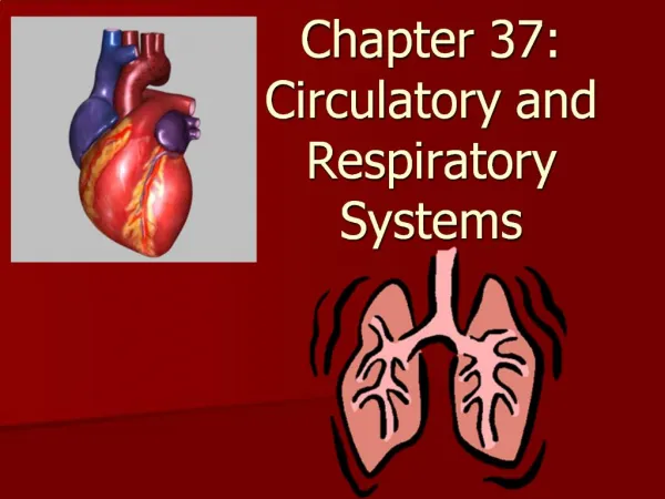
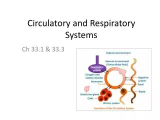
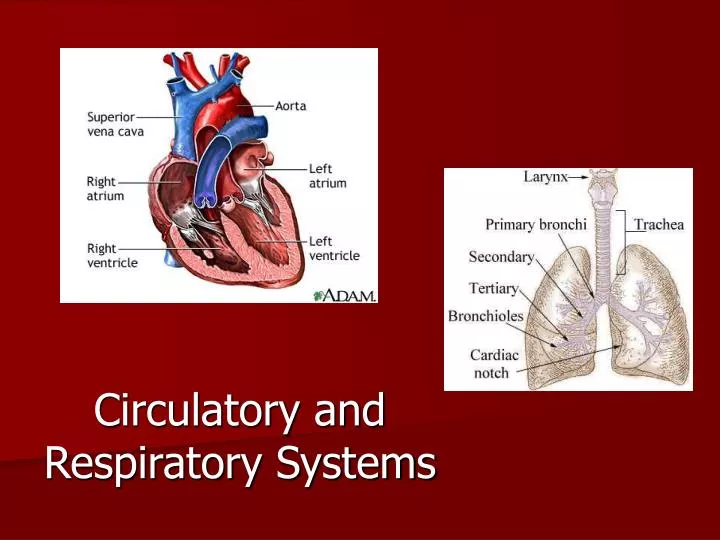
Circulatory and Respiratory Systems. Circulatory System (section 1) . Connects the muscles and organs of the body through an extensive system of vessels that transport blood, a mixture of specialized cells, and fluid. Circulatory System (section 1) .

E N D
Circulatory System (section 1) • Connects the muscles and organs of the body through an extensive system of vessels that transport blood, a mixture of specialized cells,andfluid.
Circulatory System (section 1) Different molecules the move through the cardiovascular system are: - Nutrients from digested food - Oxygen from the lungs - Metabolic wastes (carbon dioxide) - Hormones - Lastly, it distributes heat more or less evenly to maintain constant body temperature
Circulatory System (section 1) • Arteries= blood vessels that carry blood AWAY from the heart (red on diagrams) • Capillaries= tiny blood vessels that allow the exchange of gases, nutrients, hormones, and other molecules in the blood • Veins = blood vessels that carry blood BACKto the heart (blue on diagrams)
Section 37.2 Summary – pages 975-984 Your Blood Vessels: Pathways of Circulation • The three main types of blood vessels are arteries, capillaries, and veins. Aorta Right pulmonary artery (lung) Left pulmonary artery (lung) Right pulmonary veins (lungs) Capillaries in lungs Left pulmonary veins (lungs) Vena cava Heart Systemic arteries Systemic veins
Circulatory System (section 1) • A valve is a one-way flap of tissue that ensures that the blood or fluid that passes through does not flow back.
Lymphatic System • Collects and recycles fluids that leaked form the cardiovascular system and is involved in fighting infections. • Lymphtissue is located in the: spleen, thymus, tonsils, bone marrow ***Also defends the body against bacteria, virus, other infecting microbes, and cancerous cells (works with the immune system)
Plasma • About 60% of the total volume of blood is plasma (90% water/ 10% solutes)
Three types of cells in blood • Red blood cells (RBC = erythrocytes)-cells that carry oxygen. Lack nuclei and cannot make proteins or repair themselves • Hemoglobin is an iron-containing protein that binds oxygen in the lungs and transports it to the tissues of the body • Anemia = a condition in which the oxygen-carrying ability of the blood is reduced
Section 37.2 Summary – pages 975-984 Red blood cells: Oxygen carriers Side view 2.0 micrometers • The round, disk-shaped cells in blood are red blood cells. Top view 7.5 micrometers • Red blood cells carry oxygen to body cells.
Three types of cells in blood • White blood cells (leukocytes) - 1 or 2 cells for every 1,000 RBC and has a primary job to defend the body against disease (works with the immune system)
Three types of cells in blood • Platelets – cell fragments (important for clotting of blood) • Hemophilia is a blood clotting disease
Section 37.2 Summary – pages 975-984 Blood surface antigens determine blood group • Blood plasma contains proteins, called antibodies (AN tih bahd eez), that are shaped to correspond with the different blood surface antigens.
AB = Universal Recipient O = Universal Donor Rh factor = people who have this protien are said to be Rh + and those who lack it are Rh –
Section 37.2 Summary – pages 975-984 Rh factor First pregnancy • Rh factor can cause complications in some pregnancies. Placenta Rh+ antigens • Mother is exposed to Rh antigens at the birth of her Rh+ baby.
Section 37.2 Summary – pages 975-984 Rh factor • Mother makes anti-Rh+ antibodies. Possible subsequent pregnancies • During the mother’s next pregnancy, Rh antibodies can cross the placenta and endanger the fetus. Anti-Rh+ antibodies
Section 2 Check Question 2 Why is a person with type O blood considered to be a universal blood donor? (TX Obj 2; 10A)
Section 2 Check Type O blood does not contain any antigens, therefore it does not spark an immune response from the body of a person receiving the blood.
Section 2 Check Question 3 What component of blood is responsible for helping your blood clot? (TX Obj 2; 10A) A. red blood cells B. white blood cells C. plasma D. platelets The answer is D.
The Heart (section 2) • The right side of the heart is responsible for driving the pulmonary circulation loop, which pumps oxygen-poor blood through the pulmonary arteries to the lungs • The left side of the heart is responsible for driving the systemic circulation loop, which pumps oxygen-rich blood through a network of arteries to the tissues of the body.
The Heart (section 2) • Atria = are chambers that receive blood returning to the heart • Ventricles = thick walled chambers that pump blood away from the heart
Summary of Blood Flow 1. Superior Vena Cava (upper body) and Inferior Vena Cava (lower body) – sends oxygen poor blood to the right atrium 2. Right Atrium – sends blood to the right ventricle 3. Right Ventricle – sends blood to the pulmonary artery 4. Pulmonary Arteries - sends blood to the lungs to become oxygenated
Summary of Blood Flow 5. Pulmonary Veins – returns blood to the left atrium from the lungs 6. Left Atrium – sends blood to the left ventricle 7. Left Ventricle – sends blood to the aorta 8. Aorta – sends blood to the coronary arteries, the brain, and the rest of the body (sends fresh oxygen to body)
Section 37.2 Summary – pages 975-984 The passage of blood Superior vena cava Pulmonary artery Aorta LA Pulmonary vein RA LV RV Capillaries Inferior vena cava Left lung Right lung
Sinoatrial Node (SA/ Pacemaker) = contraction of the heart is initiated by a small cluster of cardiac muscle cells • Blood Pressure – is the force exerted by blood as it moves through blood vessels • 120 (systolic) / 80 (diastolic) = good reading in adults
Disorders of the Heart • Heart attack occurs when an area of the heart muscle stops working and dies ***NOTE: • Stroke is when an area of the brain dies
Section 2 Check Question 4 Why are the walls in ventricles thicker and more muscular than the walls in the atria? (TX Obj 2; 10A)
Section 2 Check Answer Superior vena cava Right lung Arch of aorta Pulmonary trunk Right atrium Left atrium Right ventricle Left ventricle Right coronary artery Rib (cut) Left lung Left coronary artery Diaphragm Cut edge of pericardium
The Respiratory System (section 3) 1. Oxygen from the outside air reaches the lungs 2. The oxygen diffuses from the alveoli to the pulmonary capillaries. At the high oxygen levels that occur in the blood within the lungs, most hemoglobin molecules carry a full load of oxygen.
The Respiratory System (section 3) 3. The oxygen-rich blood then travels to the heart. The heart pumps the blood to the tissues of the body. 4. Oxygen diffuses into the cells for use during aerobic respiration. In the tissues, oxygen levels are lower. This causes the hemoglobin to release its oxygen Hemoglobin
The Respiratory System (section 3) 5. In tissues, the presence of carbon dioxide produced by cellular respiration makes the blood more acidic and causes the hemoglobin molecules to assume a different shape, one that gives up oxygen more easily. The carbon dioxide diffuses from the cells to the blood.
The Respiratory System (section 3) 6. Most of the carbon dioxide travels to the heart as bicarbonate ions. 7. The heart pumps the blood to the lungs. In the lungs, carbon dioxide is released to its gaseous form to the alveoli. 8. The carbon dioxide is expelled.
Section 37.1 Summary – pages 971-974 The path air takes Pharynx Nasal cavity Medulla oblongata Epiglottis Larynx Esophagus Trachea Bronchus Right lung Bronchiole Left lung Diaphragm
Section 37.1 Summary – pages 971-974 Respiration and Lung Function Click image to view movie.
Diseases of the Respiratory System Asthma – a chronic condition in which the bronchioles of the lungs become inflamed because of their sensitivity to certain stimuli in the air.
Diseases of the Respiratory System Emphysema – a chronic pulmonary disease resulting from chemical imbalances that destroys elastic fibers in the lungs. Normal Emphysema
Diseases of the Respiratory System Lung Cancer – one of the leading causes of death in the world today
Two Kinds of Circulatory Systems Open Circulatory System – a heart pumps fluid containing oxygen and nutrients through a series of vessels out into the body cavity. Here the fluid washes across the body’s tissues, supplying them with oxygen and nutrients. EXAMPLE: Earthworms
Two Kinds of Circulatory Systems Closed Circulatory System – a heart pumps blood through a system of bloodvessels. Here the blood vessels form a network that permits blood flow from the heart to all of the body’s cells and back again. EXAMPLE: Humans
Section 37.2 Summary – pages 975-984 Your Blood Vessels: Pathways of Circulation • The three main types of blood vessels are arteries, capillaries, and veins. Aorta Right pulmonary artery (lung) Left pulmonary artery (lung) Right pulmonary veins (lungs) Capillaries in lungs Left pulmonary veins (lungs) Vena cava Heart Systemic arteries Systemic veins
Section 1 Check Question 5 Where does gas exchange occur during respiration? (TX Obj 2; 10A) A. in the blood B. in capillaries C. in alveoli D. in the diaphragm
Section 1 Check Alveoli The answer is C. Alveoli are the sacs of the lungs where oxygen and carbon dioxide are exchanged. O2 – rich blood Capillary network CO2 – rich blood Alveolus
Section 1 Check Question 6 How does the diaphragm enable your lungs to fill with air when you inhale? (TX Obj 2; 10A)
Section 1 Check Lung when inhaling Position of ribs when inhaling When you inhale, the diaphragm flattens, enlarging the chest cavity and drawing air into the lungs. Position of diaphragm when inhaling