Download
1 / 179
1.8k likes | 1.82k Views
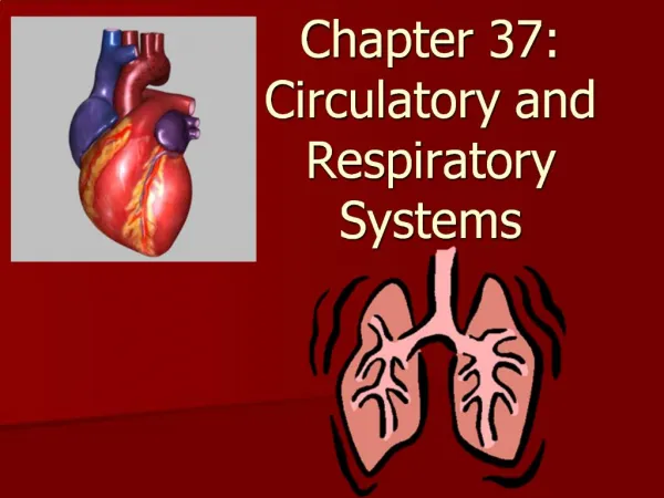
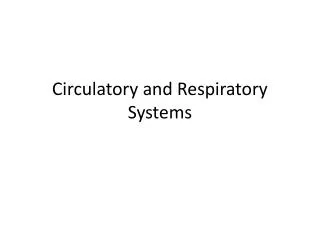
Circulatory and Respiratory Systems. This scanning electron micrograph shows individual red and white blood cells flowing through a vein (magnification 3850×). Circulatory and Respiratory Systems. The Circulatory System. Your heartbeat is a sign of life itself

E N D
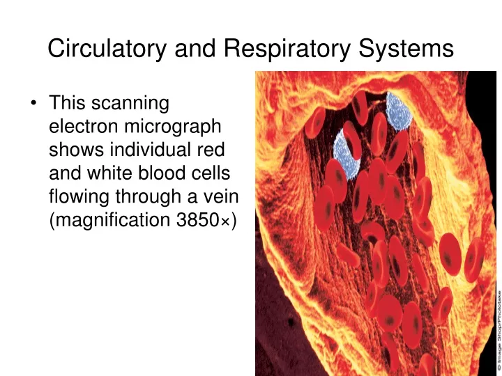
Circulatory and Respiratory Systems • This scanning electron micrograph shows individual red and white blood cells flowing through a vein (magnification 3850×)
The Circulatory System • Your heartbeat is a sign of life itself • Even when you drift off to sleep, your heart continues to beat at a steady rhythm • Why is this process so important that it must keep going even when you sleep?
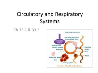
The Circulatory System • Each breath you take brings air into your respiratory system • The oxygen in that air is needed by the trillions of cells in your body • Your heart is essential in delivering that oxygen • Its beating produces the force to move oxygen-rich blood through the circulatory system • Interrelationships between the circulatory and respiratory systems supply cells throughout the body with the nutrients and oxygen they need to stay alive
Functions of the Circulatory System • Organisms composed of a small number of cellsdo not need a circulatory system • Most cells in such organisms are in direct contact with the environment • Oxygen, nutrients, and waste products can easily diffuse back and forth across cell membranes
Functions of the Circulatory System • Larger organisms, however, cannot rely on diffusion • Most of their cells are not in direct contact with the environment, and substances made in one part of the organism may be needed in another part • In a way, this same problem is faced by the millions of people living in a large city • Cities have transportation systems that move people, goods, and waste material from one place to another • The transportation system of a city is its streets, highways, and rail lines • The transportation system of a living organism is its circulatory system
CIRCULATORYSYSTEM • Consist of the heart, blood vessels, and blood • Transports gases, nutrients, hormones, and waste products throughout the body
Functions of the Circulatory System • Humans and other vertebrates have closed circulatory systems • This means that a circulating fluid called blood is contained within a system of vessels • The human circulatory system consists of the heart, a series of blood vessels, and the blood that flows through them
HEART • Muscular organ that pumps blood throughout the body • Average heart rate is 70 beats per minute • The beating noise is the opening and closing of the valves • Four chambers (2 atria/2ventricles)
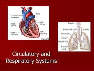
The Heart • As you can feel with your hand, your heart is located near the center of your chest • The heart, which is composed almost entirely of muscle, is a hollow organ that is about the size of your clenched fist • The heart is enclosed in a protective sac of tissue called the pericardium • In the walls of the heart, there are two thin layers of epithelial and connective tissue that form a sandwich around a thick layer of muscle called the myocardium • The powerful contractions of the myocardium pump blood through the circulatory system
HEART STRUCTURE • Pericardium: saclike membrane that surrounds the heart and secretes a fluid that reduces friction as the heart beats • Septum: divides the heart lengthwise into two pumps (right pumps deoxygenated blood to the lungs and the left pumps oxygenated blood to the body) • Atrium: thin walled upper chamber (receives blood) • Ventricle: thick walled lower chamber (pumps blood) (Which side is the thickest ?) • Blood moves from the atrium to the ventricle • One way valves (a-v valve) separate the atria and ventricles • tricuspid valve: right side • bicuspid valve: left side • pulmonary semilunar valve: exit of right ventricle • aortic semilunar valve: exit of left ventricle
The Heart • The heart muscle contracts on average 72 times a minute, pumping about 70 milliliters of blood with each contraction • This means that during one year, an average person's heart pumps more than enough blood to fill an Olympic-sized swimming pool • An Olympic-sized swimming pool is about 2,000,000 liters: 0.07 liters × 4320 beats per hour × 24 hours × 365 days = 2,649,024 liters
The Heart • Dividing the right side of the heart from the left side of the heart is the septum • The septum prevents the mixing of oxygen-poor and oxygen-rich blood • On each side of the septum are two chambers • The upper chamber, which receives the blood, is the atrium (plural: atria) • The lower chamber, which pumps blood out of the heart, is the ventricle • The heart has four chambers—two atria and two ventricles
Circulation Through the Body • The heart functions as two separate pumps • The figure shows the circulation of blood through the body • The right side of the heart pumps blood from the heart to the lungs • This pathway is known as pulmonary circulation • In the lungs, carbon dioxide leaves the blood and oxygen is absorbed • The oxygen-rich blood then flows into the left side of the heart and is pumped to the rest of the body • This pathway is called systemic circulation • Blood that returns to the right side of the heart is oxygen-poor because cells have absorbed much of the oxygen and loaded the blood with carbon dioxide • At this point, it is ready for another trip to the lungs
Circulatory Pathways • The circulatory system is divided into two pathways • Pulmonary circulation carries blood between the heart and the lungs • Systemic circulation carries blood between the heart and the rest of the body • Diagrams use red to show oxygen-rich blood, and blue to show oxygen-poor blood • What kind of blood—oxygen-rich or oxygen-poor—leaves the lungs and returns to the heart?
CIRCULATIONPATTERNS • Pulmonary Circulation • movement of deoxygenated blood from the heart (right ventricle) to the lungs by means of the pulmonary artery • blood will become oxygenated in the lungs • oxygenated blood returns to the heart (left atrium) by means of the pulmonary vein
CIRCULATIONPATTERNS • Systemic Circulation • Movement of oxygenated blood from the heart (left ventricle) to the aorta and all regions of the body by means of the major arteries • Deoxygenated blood returns to the heart (right atrium) by means of the superior/inferior vena cava • Subsystems: • Coronary: penetrates tissues of the heart • Renal: penetrates tissues of the kidneys • Hepatic portal: penetrates tissues of the liver
Circulation Through the Heart • Blood enters the heart through the right and left atria, as shown in the figure • As the heart contracts, blood flows into the ventricles and then out from the ventricles to either the body or the lungs • There are flaps of connective tissue called valves between the atria and the ventricles • Blood moving from the atria holds the valves open • When the ventricles contract, the valves close, which prevents blood from flowing back into the atria
Structures of the Heart • The circulatory system consists of the heart, a series of blood vessels, and the blood • Notice the valves between the atria and ventricles and those between the ventricles and the blood vessels leaving the heart • The valves prevent blood from flowing backward
Circulation Through the Heart • At the exits from the right and left ventricles, there are valves that prevent blood that flows out of the heart from flowing back in • This system of valves keeps blood moving through the heart in one direction, like traffic on a one-way street • The one-way flow increases the pumping efficiency of the heart • The valves are so important to heart function that surgeons often attempt to repair or replace a valve that has been damaged due to disease
Heartbeat • There are two networks of muscle fibers in the heart, one in the atria and one in the ventricles • When a single fiber in either network is stimulated, all the fibers are stimulated and the network contracts as a unit • Each contraction begins in a small group of cardiac muscle cells—the sinoatrial node—located in the right atrium • Because these cells “set the pace” for the heart as a whole by starting the wave of muscle contraction through the heart, they are also called the pacemaker
HEARTBEAT RATE • Controlled by the pacemaker (sinoatrial node: s-a node) • Located in the right atrium • Generates electrical impulse which is sent to the atrioventricular node (a-v node) located in the septum between the atria which delays the impulse about a millisecond and then sends it into the ventricles • Two nerves connect to the pacemaker • One increases the rate and the other decreases the rate
Heartbeat • As shown in the figure, the impulse spreads from the pacemaker (SA node) to the network of fibers in the atria • It is picked up by a bundle of fibers called the atrioventricular node and carried to the network of fibers in the ventricles • When the network in the atria contracts, blood in the atria flows into the ventricles • When the muscles in the ventricles contract, blood flows out of the heart • This two-step pattern of contraction makes the heart a more efficient pump
The Heartbeat • The signal to contract spreads from the sinoatrial node to the cardiac muscle cells of the atria, causing the atria to contract • The impulse is picked up by the atrioventricular node, which transmits the impulse to muscle fibers in the ventricles, causing the ventricles to contract • In times of stress, does the heart beat faster or slower?
Heartbeat • Your heart can beat faster or more slowly, depending on your body's need for oxygen-rich blood • During vigorous exercise, your heart rate may increase to about 200 beats per minute • Although the heartbeat is not directly controlled by the nervous system, the autonomic nervous system does influence heart rate • Neurotransmitters released by the sympathetic nervous system increase heart rate • Those released by the parasympathetic nervous system decrease heart rate
Blood Vessels • Blood leaving the left side of the heart is loaded with oxygen from the lungs • When it leaves the left ventricle, the blood passes into a large blood vessel known as the aorta • The aorta is the first of a series of blood vessels that carry the blood on its round trip through the body and back to the heart • As blood flows through the circulatory system, it moves through three types of blood vessels—arteries, capillaries, and veins
BLOOD VESSELS • Direction of blood flow: artery to arteriole to capillary to venule to vein • Artery: blood vessel that carries blood away from the heart • Vein: blood vessel that carries blood to the heart • Arterioles: smaller arteries • Venules: smaller veins • Capillaries: • Connect arterioles and venules • Thickness: single cell • Location of exchange between blood and body tissue
Blood Vessels • In the circulatory system, there are three types of blood vessels—arteries, capillaries, and veins • The walls of these vessels contain connective tissue, smooth muscle, and endothelium
Arteries • Large vessels that carry blood from the heart to the tissues of the body are called arteries • Arteries are the superhighways of the circulatory system • Except for the pulmonary arteries, all arteries carry oxygen-rich blood • Arteries have thick walls that help them withstand the powerful pressure produced when the heart contracts and pushes blood into the arteries • The figure shows that the walls contain connective tissue, smooth muscle, and endothelium • The elastic connective tissue allows an artery to expand under pressure • Contractions of the smooth muscle regulate the diameter of an artery
ARTERY • Same number of tissue layers as veins but thicker and more elastic • Blood under high pressure since it is coming from the heart • Tend to be deeper in the body • Arteriosclerosis: disease in which the artery walls become less elastic (blood pressure increases)
Capillaries • The smallest of the blood vessels are the capillaries • Capillaries are the side streets and alleys of the circulatory system • The walls of capillaries are only one cell thick, and most are so narrow that blood cells must pass through them in single file • The real work of the circulatory system—bringing nutrients and oxygen to the tissues and absorbing carbon dioxide and other waste products from them—is done in the capillaries
CAPILLARY • Wall consists of only a single layer of cells • Small molecules easily diffuse through this thin wall • Location of the exchange between blood and body tissues
Veins • Once blood has passed through the capillary system, it must be returned to the heart • This is the job of the veins • As with arteries, the walls of veins contain connective tissue and smooth muscle • Large veins, such as those shown in the leg in the figure below, contain valves that keep blood moving toward the heart • Many veins are located near and between skeletal muscles • When you exercise, contracting these muscles helps force blood through the veins • Blood flow through the veins of the arms and legs often occurs against the force of gravity • Exercise helps to keep blood from accumulating in the limbs and stretching the veins out of shape • If the walls around the veins weaken from lack of activity, the valves can weaken • This causes blood to pool in the veins, producing a condition known as varicose veins
VEIN • Under very low pressure • Contain valves that regulate the direction of blood flow (every few cm) toward the heart • Close if blood moves backwards • Separation of valves can result in varicose veins
Blood Vessels • Veins: Contraction of skeletal muscles helps move blood in veins toward the heart.
ARTERYBLOOD PRESSURE • Pressure results from the force the heart applies to the blood at any given time • Pressure is the highest in arteries when the ventricles contract (Systole) • Pressure is the lowest in arteries when the ventricles relax (Diastole) • Expressed as a fraction (120/80) • Systole/diastole phases create the characteristic “lubb dup” we call a heartbeat (opening/closing of valves)
HYPERTENSION • High blood pressure • Many different causes (e.g. arteriosclerosis) • Blood vessels can burst (e.g. capillaries) • Can result in kidney damage, heart attack, and even death