Download
1 / 95
950 likes | 1.05k Views
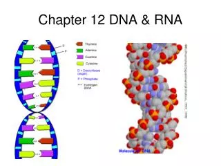
Chapter 12: D NA and RNA. KWL Chart. Learning Targets for Section 12-1. Summarize the relationship between genes and DNA? Describe the overall structure of DNA?. 12–1 Research Behind DNA Griffith and Transformation.

E N D
Learning Targets for Section 12-1 • Summarize the relationship between genes and DNA? • Describe the overall structure of DNA?
12–1 Research Behind DNA Griffith and Transformation • In 1928, British scientist Frederick Griffith tried to determine which bacteria produced pneumonia. • Griffith isolated two different strains. 1. disease-causing = smooth colonies 2. harmless strain = rough colonies.
Griffith's Experiment • Griffith injected mice 1. disease-causing bacteria- mice developed pneumonia and died. 2. harmless strain - didn’t get sick at all
Experiment cont. • Griffith’s then mixed heat-killed, disease-causing bacteria with live, harmless ones and injected the mixture into mice • Mice developed pneumonia and many died. • Found their lungs filled with the disease-causing bacteria
Figure 12–2 Griffith’s Experiment Section 12-1 Heat-killed, disease-causing bacteria (smooth colonies) Harmless bacteria (rough colonies) Harmless bacteria (rough colonies) Control(no growth) Heat-killed, disease-causing bacteria (smooth colonies) Disease-causing bacteria (smooth colonies) Dies of pneumonia Dies of pneumonia Lives Lives Live, disease-causingbacteria (smooth colonies) Go to Section:
Figure 12–2 Griffith’s Experiment Section 12-1 Heat-killed, disease-causing bacteria (smooth colonies) Harmless bacteria (rough colonies) Harmless bacteria (rough colonies) Control(no growth) Heat-killed, disease-causing bacteria (smooth colonies) Disease-causing bacteria (smooth colonies) Dies of pneumonia Dies of pneumonia Lives Lives Live, disease-causingbacteria (smooth colonies) Go to Section:
Figure 12–2 Griffith’s Experiment Section 12-1 Heat-killed, disease-causing bacteria (smooth colonies) Harmless bacteria (rough colonies) Harmless bacteria (rough colonies) Control(no growth) Heat-killed, disease-causing bacteria (smooth colonies) Disease-causing bacteria (smooth colonies) Dies of pneumonia Dies of pneumonia Lives Lives Live, disease-causingbacteria (smooth colonies) Go to Section:
Griffith’s Conclusion: • Griffith hypothesized some factor transformed harmless cells into the heat-killed harmful cells Griffith movie
Avery and DNA • Avery and his colleagues repeated Griffith’s experiment then: • Used enzymes that destroyed proteins, lipids, carbohydrates, and other molecules, including the nucleic acid RNA • When they destroyed the nucleic acid (DNA), transformation did not occur
Avery’s Conclusion: • Avery and other scientists discovered that DNA is the nucleic acid that stores and transmits the genetic information from one generation of an organism to the next
The Hershey Chase Experiment • Martha Chase and Alfred Hershey
The Hershey Chase Experiment • Studied viruses, nonliving particles smaller than a cell that can infect living organisms • Hershey and Chase reasoned that if they could determine which part of the virus—the protein coat or the DNA core—entered the infected cell, they would learn whether genes were made of protein or DNA • They grew viruses in cultures of radioactive isotopes of phosphorus-32 (32P) and sulfur-35 (35S).
The Hershey Chase Experiment • Proteins contain almost no phosphorus and DNA contains no sulfur • If 35S was found in the bacteria, it would mean that the viruses’ protein had been injected, If 32P was found in the bacteria, then it was the DNA that had been injected
Figure 12–4 Hershey-Chase Experiment Section 12-1 Bacteriophage with phosphorus-32 in DNA Phage infectsbacterium Radioactivity inside bacterium Bacteriophage with sulfur-35 in protein coat Phage infectsbacterium No radioactivity inside bacterium Go to Section:
Figure 12–4 Hershey-Chase Experiment Section 12-1 Bacteriophage with phosphorus-32 in DNA Phage infectsbacterium Radioactivity inside bacterium Bacteriophage with sulfur-35 in protein coat Phage infectsbacterium No radioactivity inside bacterium Go to Section:
Figure 12–4 Hershey-Chase Experiment Section 12-1 Bacteriophage with phosphorus-32 in DNA Phage infectsbacterium Radioactivity inside bacterium Bacteriophage with sulfur-35 in protein coat Phage infectsbacterium No radioactivity inside bacterium Go to Section:
Hershey and Chase’s Conclusion: Hershey and Chase concluded that the genetic material of the bacteriophage was DNA, not protein.
REVIEW • In your science journal: • List as many scientists you can remember • And describe their contribution to our understanding of DNA
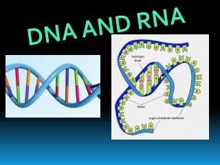
The Structure of DNA • DNA is a long molecule made up of units called nucleotides • Each nucleotide is made up of three parts: 1. a 5-carbon sugar called deoxyribose, 2. a phosphate group, 3. and a nitrogenous (nitrogen- containing) base
There are four kinds of nitrogenous bases in DNA • Purines: • Adenine: Expressed A • Guanine: Expressed G • Pyrimidines: • Thymine: Expressed T • Cytocine: Expressed C
Figure 12–5 DNA Nucleotides Section 12-1 Purines Pyrimidines Adenine Guanine Cytosine Thymine Phosphate group Deoxyribose Go to Section:
Chargaff’s Rules • Erwin Chargaff, an American biochemist, discovered that the percentages of guanine [G] and cytosine [C] are almost equal in any sample of DNA • The same thing is true for adenine [A] and thymine [T] • Despite the fact that DNA samples from organisms obeyed this rule, neither Chargaff nor anyone else had the faintest idea why
X-Ray Evidence • In the early 1950s, a British scientist named Rosalind Franklin began to study DNA using a technique called X-ray diffraction • The X-shaped pattern in the image shows that the strands in DNA are twisted around each other like the coils of a spring, a shape known as a helix
The Double Helix • At the same time Francis Crick, and James Watson, were trying to understand the structure of DNA by building three-dimensional models of the molecule • In 1953, Watson was shown a copy of Franklin’s X-ray pattern. In his book The Double Helix, Watson wrote: “The instant I saw the picture my mouth fell open and my pulse began to race.”
Watson and Crick’s Conclusion: - DNA is a double helix in which two strands are wound around each other. - Each strand is made up of a chain of nucleotides. - The two strands are held together by hydrogen bonds between adenine and thymine and between guanine and cytosine.
Learning Targets for Section 12.2 • Summarize the events that happen in DNA replication • Relate the DNA molecule to chromosome structure.
12–2 Chromosomes and DNA Replication DNA and Chromosomes Most prokaryotes have a single circular DNA molecule that contains nearly all of the cell’s genetic information
Prokaryotic Chromosome Structure Section 12-2 Chromosome E.coli bacterium Bases on the chromosome Go to Section:
Differences between Prokaryotes and Eukaryotes • Eukaryotic DNA is a bit more complicated. Many eukaryotes have as much as 1000 times the amount of DNA as prokaryotes • DNA Length DNA molecules are surprisingly long • The chromosome of the prokaryote E. coli, contains 4,639,221 base pairs
Length of DNA • This means that the nucleus of a human cell contains more than 1 meter of DNA • How can so much DNA be packed into each and every cell in our body?
Chromosome Structure • Eukaryotic chromosomes contain both DNA and protein, tightly packed together to form a substance called chromatin • Chromatin consists of DNA that is tightly coiled around proteins called histones
Chromosome Structure • Together, the DNA and histone molecules form a beadlike structure called a nucleosome • This allows the chromosomes to be very tightly coiled up in the nucleus
Figure 12-10 Chromosome Structure of Eukaryotes Section 12-2 Chromosome Nucleosome DNA double helix Coils Supercoils Histones Go to Section:
DNA Replication • When Watson and Crick discovered the double helix structure of DNA, there was one more remarkable aspect that they recognized immediately. • The structure explained how DNA could be copied, or replicated • Each strand of the DNA double helix has all the information needed to reconstruct the other half by the mechanism of base pairing
DNA Replication • During DNA replication, the DNA molecule separates into two strands • Then produces two new complementary strands following the rules of base pairing. • Each strand of the double helix of DNA serves as a template, or model, for the new strand
Figure 12–11 DNA Replication Section 12-2 Original strand DNA polymerase New strand Growth DNA polymerase Growth Replication fork Replication fork Nitrogenous bases New strand Original strand Go to Section:
DNA Replication DNA replication animation DNA Replication animation
How DNA Replicates • Start with a double strand of DNA • DNA replication is carried out by a series of enzymes. which “unzip” a molecule of DNA
How DNA Replicates A – T G – C A – T C – G T – A C – G I’ve deleted the sugar-phosphate backbone for easier drawings Hydrogen bonds