Download

1 / 76
760 likes | 924 Views
Lymphatics and the Immune System. Lymphatic System. One way system: to the heart Return of collected excess tissue fluid Return of leaked protein “Lymph” is this fluid Edema results if system blocked or surgically removed. Lymph capillaries

E N D
Lymphatic System • One way system: to the heart • Return of collected excess tissue fluid • Return of leaked protein • “Lymph” is this fluid • Edema results if system blocked or surgically removed
Lymph capillaries • Have one way minivalves allowing excess fluid to enter but not leave • Picks up bacteria and viruses as well as proteins, electrolytes and fluid (lymph nodes destroy most pathogens)
Lymph capillaries • Absent from bone, bone marrow, teeth, CNS • Enter lymphatic collecting vessels • Lymphatic collecting vessels • Similar to blood vessels (3 layers), but thin & delicate • Superficial ones in skin travel with superficial veins • Deep ones of trunk and digestive viscera travel with deep arteries • Very low pressure • Distinctive appearance on lymphangiography • Drain into lymph nodes
Lymph nodes: bean shaped organs along lymphatic collecting vessels • Up to 1 inch in size • Clusters of both deep and superficial LNs
Lymph Nodes * Superficial groups -Cervical -Axillary -Inguinal Deep groups -Tracheobronchial -Aortic -Iliac Drainage -Superior R 1/4 of body: R lymphatic duct (green) * -The rest: thoracic duct * *
Fibrous capsule sends in dividing trabeculae • Afferent & efferent lymphatic vessels • Lymph percolates through lymph sinuses • Follicles: masses of lymphoid tissue divided into outer cortex & inner medulla (details in later slides)
Macrophages on reticular fibers consume pathogens and foreign particles Usually pathogen free lymph enters lymph trunks
Lymphatic Trunks(all are paired except the intestinal trunk) • Lumbar • Intestinal • Receives fatty lymph (chyle) absorbed through lacteals infingerlike villi of intestines • Broncho-mediastinal • Subclavian • Jugular
Lymph ducts(variable) 20% • Thoracic duct: everyone has • 20% also have a right lymphatic duct *
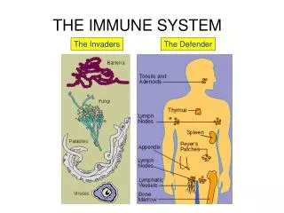
The Immune System • Recognizes specific foreign molecules • Each exposure (to the same pathogen) increases the effectivity of the response • Lymphoid organs • Lymph nodes • Spleen • Thymus • Tonsils • Small intestine & appendix aggregated lymphoid nodules
Basic Immunology • Depends on the ability of the immune system to distinguish between self and non-self molecules • Self molecules are those components of an organism's body that can be distinguished from foreign substances by the immune system • Autoimmunity is an immune reaction against self molecules (causes various diseases) • Non-self molecules are those recognized as foreign molecules • One class of non-self molecules are called antigens (short for antibody generators) and are defined as substances that bind to specific immune receptors and elicit an immune response
Lymphocytesthe primary cells of the lymphoid system • Respond to: • Invading organisms • Abnormal body cells, such as virus-infected cells or cancer cells • Foreign proteins such as the toxins released by some bacteria • Types of lymphocytes • T cells (thymus-dependent) • B cells (bone marrow-derived) • NK cells (natural killer)
T Cells • 80% of circulating lymphocytes • Some of the types: • Cytotoxic T cells: attack foreign cells or body cells infected by viruses (“cell-mediated immunity”) • Regulatory T cells: Helper T cells and suppressor T cells (control activation and activity of B cells) • Memory T cells: produced by the division of activated T cells following exposure to a particular antigen (remain on reserve, to be reactivated following later exposure to the same antigen)
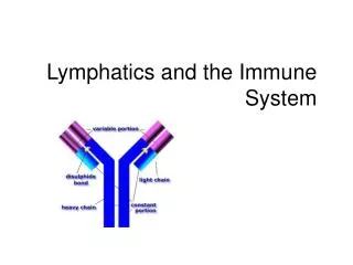
B Cells • 10-15% of circulating lymphocytes • Can differentiate into plasmocytes (plasma cells) when stimulated by exposure to an antigen • Plasma cells produce antibodies: soluble proteins which react with antigens, also known as immunoglobulins (Ig’s) • “Humoral immunity”, or antibody-mediated immunity • Memory B cells: produced by the division of activated B cells following exposure to a particular antigen (remain on reserve, to be reactivated following later exposure to the same antigen)
NK Cells • 5-10% of circulating lymphocytes • Attack foreign cells, normal cels infected with viruses, cancer cells that appear in normal tissues • Known as “immunologic surveillance”
“Humoral” vs “Cell mediated” • Cell-mediated immunity - direct attack by activated T cells (react with foreign antigens on the surface of other host cells) • Antibody-mediated (humoral) immunity – attack by circulating antibodies, also called immunoglobins (Ig’s), released by the plasma cells derived from activated B cells “humor” – from old-fashioned word for stuff in the blood, like ‘good humors’ and ‘bad humors’ These two systems interact with each other
Ab B Lymphocytes • The receptor for antigens is an antibody on B cell surface • B lymphocytes can respond to millions of foreign antigens • This capability exists before exposure to any antigens • Each lineage of B cell expresses a different antibody, so the complete set of B cell antigen receptors represent all the antibodies that the body can manufacture • A B cell identifies pathogens when antibodies on its surface bind to a specific foreign antigen • This antigen/antibody complex is taken up by the B cell and processed by proteolysis into peptides (small pieces) • As the activated B cell then begins to divide (“clonal expansion”), its offspring secrete millions of copies of the antibody that recognizes this antigen • These antibodies circulate in blood plasma and lymph, bind to pathogens expressing the antigen and mark them for destruction by complement activation or for uptake and destruction by phagocytes • Antibodies can also neutralize challenges directly, by binding to bacterial toxins or by interfering with the receptors that viruses and bacteria use to infect cells
The needs… • To be able to attack cells which have been infected • T cells target “alien” cells – they reject transplanted organs, destroy our own cells that have been infected, and kill some cancer cells: these are all treated as foreign because they have altered (antigenic) proteins on their surfaces • To be able to take care of small extracellular antigens such as bacteria which multiply outside cells, the toxins they make, etc. • Antibodies made by plasma cells (differentiated B lymphocytes) bind to antigens on bacteria, marking them for destruction by macrophages
Development of lymphocytes Originate in bone marrow from lymphoid stem cells B cells stay in bone marrow, hence “B” cells T cells mature in thymus, hence “T” cells These divide rapidly into families Each has surface receptors able to recognize one unique type of antigen= immunocompetence
Lymphocytes • Naive immunocompetent lymphocytes “seed” secondary lymphoid organs (esp. lymph nodes) • “Antigenic challenge” – full activation upon meeting and binding with specific antigen • The B cell’s antigen receptor is an antibody (see slide 20) • Full activation • Gains ability to attack its antigen • Proliferates rapidly producing mature lymphocytes • Mature lymphocytes re-circulate seeking same pathogens
Innate immune system Response is non-specific Exposure leads to immediate maximal response Cell-mediated and humoral components No immunological memory Found in nearly all forms of life (plants & animals) Adaptive immune system Pathogen and antigen specific response Lag time between exposure and maximal response Cell-mediated and humoral components Exposure leads to immunologic memory Found only in jawed vertebrates Components of the immune system
Innate immunity • The dominant system of host defense in most organisms • Inflammation is one of the first responses • Redness, swelling, heat and pain • Chemical and cellular response • During the acute phase of inflammation, particularly as a result of bacterial infection, neutrophils migrate toward the site of inflammation in a process called chemotaxis, and are usually the first cells to arrive at the scene of infection
Lymphoid Organs • Lymph nodes • Spleen • Thymus • Tonsils • Small intestine & appendix aggregated lymphoid nodules
Lymphoid Tissue Specialized connective tissue with vast quantities of lymphocytes • Lymphocytes become activated • Memory • Macrophages & dentritic cells also • Clusters of lymphoid nodules or follicles
Thymus • Prominent in newborns, almost disappears by old age • Function: T lymphocyte maturation (immunocompetence) • Has no follicles because no B cells
Lymph Nodes • Lymphatic and immune systems intersect • Masses of lymphoid tissue between lymph sinuses (see next slide) • Some of antigens leak out of lymph into lymphoid tissue • Antigens destroyed and B and T lymphocytes are activated: memory (aiding long-term immunity)
Follicles: masses of lymphoid tissue divided into outer cortex & inner medulla • All follicles and most B cells: outer cortex • Deeper cortex: T cells, especially helper T cells • Medullary cords: T & B lymphocytes and plasma cells
Spleen • Largest lymphoid tissue; is in LUQ posterior to stomach • Functions • Removal of blood-borne antigens: “white pulp” • Removal & destruction of aged or defective blood cells: “red pulp” • Stores platelets • In fetus: site of hematopoiesis • Susceptible to injury; splenectomy increases risk of bacterial infection
Tonsils Simplest lymphoid tissue: swellings of mucosa, form a circle Crypts get infected in childhood Palatine (usual tonsillitis) Lingual (tongue) Pharyngeal (“adenoids”) Tubal * * *
Parts of the intestine are so densely packed with MALT (mucosa-associated lymphoid tissue) that they are considered lymphoid organs • Aggregated lymphoid nodules (“Peyer’s Patches”) • About 40 follicles, 1 cm wide • Distal small intestine (ileum) • Appendix
The Lymphatic System and Our immune response: Did you know? • Laughing lowers levels of stress hormones and strengthens the immune system. Six-year-olds laugh an average of 300 times a day. Adults only laugh 15 to 100 times a day. • 3000 BC The ancient Egyptians recognize the relationship between exposure to disease and immunity. • 1500 BC The Turks introduce a form of vaccination called variolation, inducing a mild illness that protects against more serious disease. • 1720 Lady Mary Wortley Montagu promotes the variolation principle, launching a campaign to inoculate the English against smallpox. • A macrophage can consume as many as 100 bacteria before undergoing apoptosis.
What does the lymphatic system do? • Return interstitial fluid • Capillaries only reabsorb 15% • Funneled into subclavian veins • Absorb and transport lipids from intestines • Generate and monitor immune responses • lymphatic system movie
What is in the lymphatic system? • Lacteals and lymphatic capillaries • Overlapping epithelial cells • Lymph vessels and ducts • What happens if blockage occurs? • See next slide!
What is in the lymphatic system? • Lymphatic trunks • Lumbar, brachiomediastinal, intestinal, jugular, subclavian, intercostal • R lymphatic duct: R arm, R thorax, R head • Thoracic duct: everything else
What is in the lymphatic system? • Red bone marrow • Hemopoiesis: what types of leukocytes are manufactured here? • Mucosa-associated lymphatic tissue • Sprinkling of lymphocytes in mucosa membranes • Peyer’s patch: small intestine nodules of lymphatic tissue
What is in the lymphatic system? • Thymus • Secretes thymopoietin for T-cell development • T-cells mature here • Thymus atrophies with age • Tonsils • Palatine (2), lingual (2), pharyngeal (1; adenoid) • Tonsillectomy: remove palatines • Gather, remove and “learn” pathogens from food/air • Calculate risk in childhood: inviting invasion • Payoff: greater immunocompentency later in life
What is in the lymphatic system? • Lymph nodes • Filters lymph fluid for antigens, bacteria, etc. • B-lymphocytes made here • Some T-lymphocytes and macrophages congregate • Afferent (more) and efferent (less) vessels • lymph fluid exits through hilum • Common site for cancer—Why? • Hodgkin’s lymphoma: lymph node malignancy • Etiology unknown • Non-Hodgkin’s lymphoma: all other cancers of lymphoid tissue • Multiplication/metastasis of lymphocytes • 5th most common cancer
What is in the lymphatic system? • Spleen: dense sieve of reticular CT • Functions • Erythropoiesis in fetus • Stores platelets • Salvages and stores RBCs parts for recycling (RBC graveyard) • Red pulp • Dispose of damaged/dead RBCs and pathogens • Old RBCs aren’t flexible enough to get through sieve • White pulp • Lymphocytes and macrophages • B-cells proliferate here • If splenectomy: liver and marrow take over duties
What’s behind non-specific immunity? • Phagocytes • Macrophages: tissue-living monocytes • Neutrophils: digestion and killing zone (H2O2; superoxide ion and hypochlorite (bleach)) • Eosinophils: less avid digesters • Basophils and mast cells: it mobilize other WBCs (via histamine and heparin) • some phagocytosis • Natural Killer cells (NK cells): type of T-cell • Only attack infected or cancerous host cells
What’s behind non-specific immunity? • Inflammation • Redness, swelling, heat, pain • Bradykinin: pain stimuli from mast cells • Histamine: what two things does it do? • Leukocyte migration • Margination • http://www.med.ucalgary.ca/webs/kubeslab/home/ • Diapedesis: http://www.constantinestudios.com/animation4.html • Chemotaxis • Phagocytosis
What’s behind non-specific immunity? • Interferons • Virus-infected cells secrete warning (click here or on movie to right) • Can promote cancer cell destruction • Complement proteins • 20+ beta-globulins which perforate bacterial cells (cytolysis) • complement movie • Fever (pyrexia) • Promotes interferon activity • Elevates BMR • Discourages bacteria/viral reproduction • fever movie