Download
1 / 26
260 likes | 294 Views
Orbital Disorders 1. By Professor Ahmad Mostafa. The following will be discussed in the following order:. Applied orbital Anatomy Proptosis Orbital Infections & Inflammations Orbital Trauma (Blow-out fracture). Clinical Anatomy.

E N D
Orbital Disorders1 By Professor Ahmad Mostafa
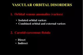
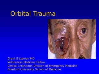
The following will be discussed in the following order: • Applied orbital Anatomy • Proptosis • Orbital Infections & Inflammations • Orbital Trauma (Blow-out fracture)
The orbit is a pear-shaped cavity whose stalk is the optic canal
● The medial walls of the two orbits are parallel, and the lateral walls form an angle of almost 90 degrees with one another.
Orbital Anatomy includes: • Orbital Dimensions • Bony Orbit (C) Orbital Contents
(A) Orbital Dimensions • The widest orbital dimension is 1 cm behind the orbital rim. • The volume of the orbit is 30 ml. • The intraorbital portion of the optic nerve is longer (25 mm) than the distance between the back of the globe and the optic foramen (18 mm).This allows for significant forward displacement of the globe (proptosis) without causing excessive stretching of the optic nerve.
(B) Bony Orbit 1. Orbital Walls 2. Orbital Foramina
(1) Orbital Walls ■ Seven bones contribute to the orbital walls ■ These are: frontal, sphenoid, zygomatic, palatine, maxillary, ethmoid, and lacrimal bones.
►Each orbit has the following 4 walls: • Orbital roof • Medial wall • Orbital floor • Lateral wall
1. Orbital Roof • It consists of 2 bones: the lesser wing of sphenoid and frontal bones.
Clinically important landmarks • It is located adjacent to the anterior cranial fossa and frontal sinus. 2. The roof contains two important fossae; the lacrimal gland fossa which is situated laterally, and the fossa for the trochlea which is found medially, approximately 4.0 - 5.0 mm posterior to the superior orbital rim. 3. A defect in the orbital roof may cause pulsating exophthalmos as a result of transmission of CSF pulsations to the globe through the defect. This defect may be either congenital (as in neurofibromatosis), or traumatic.
2. Orbital medial wall • It consists of 4 bones: maxillary, lacrimal, ethmoid,and sphenoid.
Clinically important landmarks 1. The medial wall is the thinnest of the orbital walls. 2. The lamina papyracea is a very thin plate of bone in the medial wall of the orbit which separates the ethmoid sinus from the orbital cavity. It is perforated by numerous foramina for nerves and blood vessels. Therefore, The most common cause of orbital cellulitis in children is ethmoidal sinusitis.
3. Orbital floor • It consists of 3 bones: zygomatic, maxillary, and palatine.
Clinically important landmarks • The floor of the orbit forms the roof of the maxillary sinus, so, maxillary tumors e.g. maxillary carcinoma may invade the orbit and displace the globe upwards. 2. The posteromedial portion of the maxillary bone is relatively weak, so, easily involved in blow-out fracture of the orbital floor. 3. The inferior rectus muscle is in close contact with the floor posteriorly. It can be easily damaged during transantral decompression of the orbital floor in dysthyroid ophthalmopathy.
4. Orbital lateral wall • It consists of 2 bones: the greater wing of sphenoid and zygomatic.
Clinically important landmarks 1. It is the thickest wall, so, it is not common to be involved in blow-out fracture of the orbit. 2. The anterior half of the lateral orbital wall is vulnerable to lateral trauma because it protects only the posterior half of the globe.
(2) Orbital Foramina • There are several openings (foramina) in the orbital bones that are of clinical importance. • These include the optic canal, superior orbital fissure, inferior orbital fissure, the ethmoidal foramina, and the zygomatico-facial and zygomaticotemporal foramina.
Optic nerve foramen(Optic canal) • It is formed by 2 roots of the lesser wing of sphenoid bone. • It transmits: - Optic nerve - Ophthalmic artery - Sympathetic nerves ● Evidence of enlargement of the optic canal on x-ray can be seen with optic nerve tumors.
Superior orbital fissure • It is situated between the greater and lesser wings of sphenoid bones (between lateral wall & roof of the orbit) at the orbital apex. • It connects the orbit with MCF. • It transmits: • 3rd, 4th, 6th cranial nerves • Ophthalmic branch of trigeminal (5th) nerve • Superior ophthalmic vein • Some sympathetic nerve fibres.
Acute idiopathic inflammation in the region of the superior orbital fissure produces painful ophthalmoplegia known as Tolosa-Hunt syndrome.
Inferior orbital fissure • It lies between the greater wing of sphenoid and maxilla (between lateral wall & floor of the orbit). • Through it, the orbit communicates with the pterygo-palatine and infra-temporal fossae. • It transmits: - Infraorbital nerve and artery - Zygomatic nerve - A communication between the inferior ophthalmic veins and pterygoid plexus
(C) Orbital Contents • The globe • Extraocular muscles • Orbital blood vessels (arteries & veins) • Peripheral nerves including 3rd, 4th, 5th, 6th, and sympathetic & parasympathetic fibers. • Optic nerve and meninges • Adipose tissue • Lacrimal gland