Download
1 / 26
530 likes | 1.32k Views
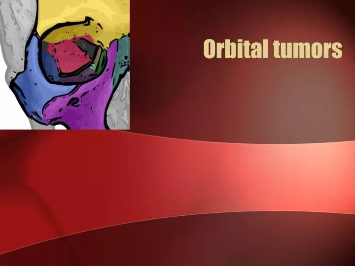
Orbital tumors. Clinical evaluation. “ Six p ’ s ” of orbital lesions: Proptosis Pain Progression Pulsation Palpation Periorbital changes. Surgical indications. Biopsy Lesions affecting vision Lesions affecting the globe Compression of the optic nerve. Complications .

E N D
Clinical evaluation “Six p’s” of orbital lesions: • Proptosis • Pain • Progression • Pulsation • Palpation • Periorbital changes
Surgical indications • Biopsy • Lesions affecting vision • Lesions affecting the globe • Compression of the optic nerve
Complications • PTOSIS: levator muscle & / or its nerve damage • DIPLOPIA : EOM damage, ocular motor nerve damage, adhesions of EOM, trochlea damage • VISUAL LOSS: CRA trauma / occlusion, globe compression, optic nerve trauma / compression (Hemorhage, edema) • CSF LEAK: inadvertent opening of the paranasal sinuses(post ethmoid ) while optic canal deroofing. • EYELID MALPOSITION: faulty wound closure, adhesions b/w lids & orbital rim • PUPIL & ACCOMODATION ABNORMALITIES: Posterior ciliary N & vessels damage
PULSATING PROPTOSIS: Due to extensive deroofing of the orbit • FRONTAL BRANCH OF FACIAL NERVE INJURY: Incision >4cm from the lateral canthal margin in lateral orbitotomy • OCULAR OR FACIAL SENSORY LOSS: sensory nerve damage (nasociliary N, 1st/2nd division of trigeminal N) • CORNEAL ULCERATION: direct corneal trauma, corneal dessication
Classification • Primary / Secondary / Metastatic • Intraconal / Extraconal / Intracanalicular • PATHOLOGICAL • Cystic : dermoid / epidermoid • Vascular : hemangioma / lymphangioma • Inflammatory : pseudotumor • Infiltrating : lymphoid tumors / LCH • Mesodermal : Fibroma/lipoma • Neurogenic: glioma / meningioma • Lacrimal : adenoma / carcinoma • Metastatic : Neuroblastoma/ Ewings • Intraocular : Retinoblastoma
Cystic lesions • The most common space-occupying masses in the orbit, representing 30% - 46% of excised orbital tumors in children • Frequently located anterior to the orbital septum along the fronto-zygomatic suture • Small cysts: close observation • Large cysts: excision in toto
Vascular lesions • Approximately 15% of cases in several series • Capillary hemangioma • MC vascular orbital tumor in cildhood • Spontaneous involution • Vision preservation dictates management • Observation/Steroids/ Co2 laser/interferon alpha • Cavernous hemangioma • Adults • Well circumscribed • Surgical excision • Lymphangioma • 1-3 % • Slowly progressive • Soft bluish mass superonasal quadrant • Bleeding – chocolate cyst • Steroids / surgical debulking
Neurogenic tumors • Gliomas • Meningioma • Neurofibroma • Schwannoma • Esthesioneuroblastoma • Paraganglioma • Melanotic neuroectodermal tumor of infancy
Optic nerve sheath meningioma • 2% of all orbital tumors and 1–2% of all meningiomas. • Primary ONSM: • 92% intraorbital nerve sheath • 8% are intracanalicular in origin. • Bilateral and multifocal presentations : NF2 • Presentation : Triad : visual loss/optic atrophy/opticociliary shunts
Management • Recommendations for observation without treatment should be followed only with caution • Surgery : • Functional vision significantly compromised • Disfiguring proptosis • Intracranial extension • Stereotactic fractionated radiotherapy : better visual outcome • Chemotherapy : Unresectable / Recurrent/ Post RT • 5 FU, Folate, levamisole
Optic nerve Gliomas • 3-5% of childhood brain tumors. 11-30% with NF1 • Typically occurs in the first decade of life • Optic disc and nerve 25%, chiasm 40–75% • Presentation : vision loss/ proptosis / strabismus / endocrinopathy • Histologically : (LGG) pilocytic / fibrillary / pilomyxoid astrocytoma • Biopsy only if unusual clinical / imaging findings.
Imaging • An enlargement of the optic nerve without calcification, as tubular / fusiform / lobulated • Classically a J-shaped sella • Optic foramina views : optic foramen > 7.0 mm or a difference of more than 2.0 mm. • T2WI demonstrate homogeneous high signal intensity of the affected nerve in contrast to the low signal of the C/L unaffected optic nerve
MANAGEMENT • Observation: newly diagnosed OPG • Surgery : Single nerve with disfiguring proptosis / blindness Exophytic chiasm tumor with hydrocephalus / ME • Chemotherapy : 1st line for symptomatic OPG beyond observation • Packer regimen: concurrent carboplatin and vincristine • Radiation therapy: progressive chiasmatic tumors in > 10 yr age , 45 – 50 Gy Optic pathway gliomas : a review Neurosurg Focus 23 (5):E2, 2007
PROGNOSIS • Confined to optic nerve: • Treated : 0% tumor-related mortality. • Observed: 21% exhibited progression 5% died 91% maintained stable vision. • Chiasmatic gliomas : 42% rate of progression / recurrence 29 % Tumor related mortality. • Good prognosis: NF1 and anterior location • Poor prognosis: younger age at presentation
Peripheral nerve tumors • Constitute 5 –15 % of the orbital tumors. • 5 types : Solitary neurofibroma Diffuse neurofibroma Plexiform neurofibroma Schwannomas Malignant peripheral nerve tumors
Solitary neurofibroma • B/w 3rd – 4th decade • Slowly progressive, painless proptosis with minimal or no visual dysfunction • Typically located in the superolateral orbital quadrant • Isointense to brain & muscle on T1WI & hyperintense to fat onT2WI with heterogenous enhancement • Pseudocapsule : easy to dissect • Prognosis is good • No need for postoperative RT.
Plexiform neurofibroma • Associated with NF • Occur mostly in infants & children • A palpable mass in the eyelid (usually lateral third) with subsequent ptosis & lid hypertrophy • May spread to forehead or adjacent areas of temple
Schwannomas(Neurilemmomas, Neurinoma) • 2nd – 5th decade, F>M • Usually originate from sensory branch of the trigeminal nerve • High incidence in patients with NF 2. • Well encapsulated • C/f : Proptosis, trigeminal distribution of pain & numbness • T1WI : Iso- to hypointense signal in relation to the orbital fat with a varying degree of contrast enhancement • Malignant transformation is rare.
Metastatic Tumors • 8% of all orbital tumors • Most common in women – breast • Most common in men – prostate & lung • Symptoms – proptosis, diplopia, pain, vision loss • Presents in 7th decade • FNAB for diagnosis (80%) • Prognosis is very poor (avg. survival 10 months) • XRT usual; Chemo and Hormonal occasional
FIBROUS DYSPLASIA • Normal cancellous bone is replaced by immature woven bone and fibrous tissue. • 2.5% of all bone tumors. • Frontal, sphenoid, ethmoid, and maxillary bone complexes • Sclerotic/ cystic (lytic)/ mixed varieties ( 40% )of cases. • classic “ground-glass” appearance on CT. • Surgery: cosmetic deformity / progressive vision loss
AIIMS Data Search • 50 Orbital tumors • Haemangioma: 11, Lymphangioma :2 • Meningioma 6 : ONSM 2 • Pseudotumor: 5 • Lacrimal gland tumor: 5 • Dermoids :3, mucocele: 1 • Metastatic: 3 • Glioma/ haemangiopericytoma/chondrosarcoma / fibrous dysplasia : 2 each • GCT/ABC/Amyloidosis :1 each From 2009 OCT– 2011 OCT