Download
1 / 20
350 likes | 658 Views
The Digestive System. Megan Malach. The structures:. Mouth Salivary glands Esophagus Upper esophageal sphincter Liver Stomach Pancreas. Gallbladder Lower esophageal sphincter Duodenum Small intestine Ileum Jejunum Large intestine Colon Anus. Mouth.

E N D
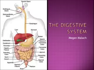
The Digestive System Megan Malach
The structures: • Mouth • Salivary glands • Esophagus • Upper esophageal sphincter • Liver • Stomach • Pancreas Gallbladder Lower esophageal sphincter Duodenum Small intestineIleumJejunum Large intestineColon Anus
Mouth • Chewing of food and digestion of starch • Contains salivary glands, tongue, and teeth in order to carry out this purpose • Food is crushed so that it may be passed along the esophagus and into the stomach Mouth
SALIVARY GLANDS • Produce salivary amylase, which initiates the breakdown of polysaccharides to simpler carbohydrates • Moistens food so that it can be chewed and swallowed • Saliva also helps to clean the mouth and kill germs
ESOPHAGUS • “Tube” that transfers food from mouth to stomach • When food enters, causes contractions of muscle • Contractions known as “peristalsis”, this is how food is moved down to stomach • Peristalsis= why you can swallow upside down This kid is trying to swallow upside down
Lower esophageal sphincter • Sphincter “above” stomach and “below” esophagus • When contracted, food cannot enter the stomach When open, food may pass
STOMACH • Storage of food and initial digestion of proteins • Secretes hydrochloric acid (HCL) (breaks down food as it is highly acidic), pesinogen (enzyme), and mucus
Liver • Secretes bile (helps with the breakdown of fats in stomach), emulsifies fat • Bile helps move fat along, and helps absorb fat soluble vitamins (vitamins “trapped” in fat) • Liver controls use of carbohydrates, lipids, and proteins • Largest organ in body This is liver pate (I think there’s an accent on the e). It’s not actually as bad as it sounds...
Pancreas • Production of digestive enzymes that act on food when it reaches the small intestine, stores bicarbonate ions to nuetralize HCL in stomach • Secretes pancreatic amylase (helps to break down carbohydrates), bicarbonate ions (to nuetralize HCL before it reaches the small intestine), trypsinogen, and lipase
GALLBLADDER • Stores and secretes concentrated bile from the liver • Can hold 50 mL of bile • Shaped like a pear • Comes in plush form!
Pyloric EsophagealSPhincter • Muscle that helps control movement of food • Sphincter “below” stomach and “above” small intestine • When contracted, food cannot enter stomach. When open, food can enter stomach The red thingie is where the lower sphincter is. This one’s here.
Duodenum • Passage between stomach and intestines • Where food moves after mixing with stomach acid • Initial absorption of vitamins, minerals, other nutrients, etc begins in duodenum • Here, food mixes with bile from the gallbladder and digestive juices from pancreas (amylase, bicarbonate ions, etc)
SMALL INTESTINE • Digestion of carbohydrates, proteins, lipids; the absorption of nutrients • Composed of iluem (lower half) and jejunum (upper half)
JEJUNUM • Vitamins A, D, sodium, water, and bile salts absorbed here • Most vitamins and minerals absorbed into body than at any other stage of digestion • Directly follows the duodenum
ILEUM • Where any remaining nutrients from breakdown of protein (and some water-soluble vitamins) absorbed • Last part of ileum important, only place where vitamin B12 can be absorbed
Large intestine • Absorption of water and storage of undigested food • Composed of three sections: ascending colon, traverse colon, and descending colon • Secretes mucus • Produces solid waste
COLON • Absorbs remaining water and electrolytes from indigestible food • Absorbs fibre (which is technically indigestible) • Keeps body’s fluids regulated • Forms and helps to dispose of feces
Anus (You don’t want to see the pictures on google for this one. Trust me.) • Connected to rectum (empties colon of feces) • Contains muscles (sphincters, internal and external) that are always contracted, so that feces only escape the body at an appropriate time • The end of the digestive system
Bibliography • http://www.vitallywell.net/images/digestive-system-diagram.jpg • http://www.nlm.nih.gov/medlineplus/ency/article/002347.htm • http://vsearch.nlm.nih.gov/vivisimo/cgi-bin/query-meta?v%3Aproject=medlineplus&query=mouth&x=0&y=0 • http://mset.rst2.edu/portfolios/d/demarco_a/toolsdev/finpro/s02osms.htm • http://2.bp.blogspot.com/_Nlofy7L_4ZE/TBMvCqKWrqI/AAAAAAAADnc/jA_RSVPyiJI/s1600/IMG_9068.JPG • http://www.vivo.colostate.edu/hbooks/pathphys/digestion/pregastric/salivary.html • http://www.vivo.colostate.edu/hbooks/pathphys/digestion/liver/bile.html • http://www.vivo.colostate.edu/hbooks/pathphys/digestion/stomach/pepsin.html • http://www.ece.ncsu.edu/imaging/MedImg/SIMS/GE1_3.html • http://www.eatatease.com/jejunum.html • http://www.eatatease.com/ileum.html • http://www.puristat.com/coloncleansing/colonfunction.aspx • http://www.cchs.net/health/health-info/docs/1600/1699.asp?index=7041 • http://alpinesurgical.net/uploads/expertise/esophagus.jpg
More Bibliography • http://www.thefatlossauthority.com/fat_loss_tips/wp-content/uploads/2010/11/Liver-pate2.jpg • http://upload.wikimedia.org/wikipedia/commons/thumb/2/29/Regions_of_stomach.svg/300px-Regions_of_stomach.svg.png • http://images.mylot.com/userImages/images/postphotos/2107804.jpg • http://static.neatoshop.com/images/product/42/942/Gallbladder-Plush_3667-l.jpg • http://www.drbhandari.com/images/esophagus.gif • http://s3.hubimg.com/u/2373094_f260.jpg • http://lh3.ggpht.com/_eXgTWwvGr0A/SoOnuUIe0BI/AAAAAAAAAYI/cHjzhWyRWxU/SmBowelAnat%5B3%5D.jpg • http://dirtpoormedic.files.wordpress.com/2011/03/large-intestines.png • http://3.bp.blogspot.com/_E8BpJEni77I/S5kMZvbZLLI/AAAAAAAAL6o/CuRAPSA-WB0/s400/colonblow_2.png


