Download
1 / 23
230 likes | 235 Views
An improved deep learning approach for detection of thyroid papillary cancer in ultrasound images. Li et al, 2018. Outline. Background Methods Results. Background. Object detection : classification + localization Classifcation : what is the object ?

E N D
An improved deep learning approach for detection of thyroid papillary cancer in ultrasound images Li et al, 2018
Outline • Background • Methods • Results
Background • Object detection: classification + localization • Classifcation: whatis the object? • Localization: whereis the object (x, y, w, h)? Krizhevsky et al, ImageNet classification with deep convolutional neural networks, 2012
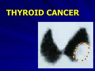
Background • Ultrasound images are monochrome and low-resolution. • Cancer regions are usually blurred, vague margin and irregular in shape
Background • CNN (breast cancer MR image) • fine-tuning • de-noising auto-encoders • CSFaster R-CNN • add a spatial constrained layer to Faster R-CNN • concatenating the shallow and deep layers of CNN
Methods • R-CNN: Regions with CNN • SPP Net: Spatial Pyramid Pooling • Fast R-CNN • Faster R-CNN • CS Faster R-CNN
R-CNN • 2000 Region Proposal: Selective Search • Warped region • CNN, feature extraction • SVM classification • Bounding-box regression Girshick et al, Rich feature hierarchies for accurate object detection and semantic segmentation, 2014
R-CNN • CNN, feature extraction • fine-tuning : (N + 1)-way classification layer • region proposals with ≥ 0.5 IoU overlap with a ground-truth box as positives for that box’s class, else negatives • Object category classifiers 21 SVM • Bounding-box regression • predict a new bounding box for the detection • G = (Gx,Gy,Gw,Gh)
SPP Net • In R-CNN warped region may result in unwanted geometric distortion • Convolutional layers do not require a fixed image size. Fully-connected layers need to have fixed size input. • Spatial Pyramid Pooling Layer He et al, Spatial Pyramid Pooling in Deep Convolutional Networks for Visual Recognition, 2015
Fast R-CNN • Input: images and a list of region of interest (RoIs) • Use a RoI pooling layer extracts a fixed-length feature • Output layers: 1 use softmax probability estimates (K+1) object; 2 outputs four real-valued numbers for each of the K object classes. • Multi-task loss: Girshick et al, Fast R-CNN, 2015
Faster R-CNN • In R-CNN, 2000 Region Proposal • Insert Region Proposal Network (RPN) to predict proposals from features • 4 Loss functions: • RPN calssification(anchor good.bad) • RPN regression(anchor->propoasal) • Fast R-CNN classification(over classes) • Fast R-CNN regression(proposal ->box) Ren et al, Faster R-CNN: Towards Real-Time Object Detection with Region Proposal Networks, 2015
Faster R-CNN • RPN • slide a small network over the convolutional feature map output by the last shared convolutional layer • at each location generate 9 anchors(3 scales and 3 aspect ratios ) • This architecture is implemented with an n×n convolutional layer followed by two sibling 1 × 1 convolutional layers (for reg and cls, respectively). • Loss function:
CS Faster R-CNN • ZF modle • Concatenate conv3 layer and conv5 layer • Spatial constrained layer • cancer regions depend on their residing regions which are hard to define.
CS Faster R-CNN • 300 cases, 4670 ultrasound images • 200 diagnosed cases as training samples • 50 diagnosed cases and 50 normal cases as test samples • CNN pre-trained ZF model with VOC2007 database
Results • ID1 does not use any strategy. ID2 uses the strategy of layer concatenation. ID3 uses the strategy of layer concatenation and spatial constrained layer.
Discussion • Small amount data, ultrasound images are limited and difficult to obtain. • fine-tuning a CNN that has been pre-trained • Ultrasound images are usually blur, vague margin, irregular shape • identify their local texture features, layer concatenation • It is difficult to identify the boundaries of cancer