Download
1 / 45
470 likes | 670 Views
Formation and patterning of the nervous system. I. Neural Induction and Neurulation - specification of neural fate and formation of the neural tube. II. Neural Patterning - patterning of neural progenitors along the dorsoventral axis

E N D
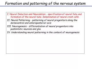
Formation and patterning of the nervoussystem I. Neural Induction and Neurulation - specification of neural fate and formation of the neural tube. II. Neural Patterning - patterning of neural progenitors along the dorsoventral axis III. Neurogenesis - differentiation of neural progenitors into postmitotic neurons and glia. IV. Understanding neural patterning in the context of neurogenesis
Organization of the spinal cord Postmitotic neurons in “mantle” layer Progenitors (dividing) in ventricular zone Fig. 11.12
The DNA content of a cell reveals its position in the cell cycle Cells in S phase can be detected by their ability to incorporate labeled DNA precursors whose presence can be detected after labeling the cells (i.e BrDU or 3H labeling - detection of proliferating cells in the CNS, birthdating neurons, cell fate).
Neurons are generated from mitotically active precursors and different types of neurons are generated in an orderly progression VZ = ventricular zone IZ = intermediate zone PP = preplate SVG = subventricular zone SP = subplate CP = cortical plate MZ = marginal zone
The DNA analogs 3H-thymidine and Bromodeoxyuridine (BrdU) can be used to “birthdate” neurons Cells that exit the cell cycle after labeling will be heavily labeled; cells that divide again dilute out the label Incorporated into DNA during S-phase, but only available for a short time (then metabolized)
Cortical neurons are generated in an inside-first, outside-last order
What possible mechanisms might control the fates of cells produced at distinct times during development? In the ferret layer6 cells are born in utero, weeks later cells are fated for layers 2 and 3 and must migrate through layers 6, 5 and 4 which are already formed. Are cortical cells determined with respect to layer as they are generated or is their fate dictated from the position they migrate to? How could one test these possibilities experimentally?
Possible outcomes Cells are multipotent; environment determines fate Cells are committed to a specific fate prior to migration
The behavior of early progenitors depends on their stage in the cell cycle at the time of transplantation 1. Cells transplanted in S-phase change fates and become normal layer 2/3 neurons
The behavior of early progenitors depends on their stage in the cell cycle at the time of transplantation 1. Cells transplanted in S-phase change fates and become normal layer 2/3. 2. Cells transplanted after the S- to-G2 transition are committed layer 6 neurons
Early progenitors are multipotent; what about late progenitors?
Late progenitors are intrinsically and heritably committed to generating upper-layer neurons Transplant progenitors from older embryos into younger brains:
What molecular mechanisms might control changes in progenitor cells over time? 1. First need to make more progenitors 2. To start to make neurons
What molecular mechanisms might control changes in progenitor cells over time? 2 G 3 G …and glia! G 4 G G 5 6
Each neuroblast emerges through a series of cell-cell interactions and changes in gene expression.
Identification of “proneural” mutations and Ac-Sc bHLH factors Schematic representation of the structure of a bHLH dimer that is complexed to DNA. The basic region fits in the main groove of the DNA, and many residues in this region make direct contact with the E-box sequence. The two -helices of both partners together form a four-helix bundle
Identification of “proneural” mutations and Ac-Sc bHLH factors Models of interactions of proneural proteins with cofactors that confer functional specificity. Functional specificities among proneural proteins. The functional specificities of the Drosophila proteins Scute (Sc) and Atonal, which are proneural factors for external sense organs and chordotonal organs, respectively, reside in residues that are located in the basic domain34. Residues that differ between the basic regions of Scute and Atonal are predicted not to directly contact the DNA, but to be involved in interactions with cofactors. In this model, a cofactor interacts with both the basic motif of the proneural protein and the DNA sequence, and provides the proneural protein with specificity for binding to a particular E-box sequence
Each neuroblast emerges through a series of cell-cell interactions and changes in gene expression.
Neurogenic genes in Drosophila: Notch, Delta, Enhancer of Split Schematic illustration of the Notch and Delta genes products: Notch encodes a large transmembrane protein 300kD,that contains a large cytoplasmic domain, a single membrane-spanning segment and a large amino-terminal extracellular domain ( a receptor). Delta, encodes a smaller homologue of Notch with nine-EGF-like repeats in the extracellular domain. Genetic experiments suggested that Notch and Delta interact with one another and biochemical experiments showed that this interaction was direct. Enhancer of Split, encodes for bHLH transcription factors, transcriptional readout of Notch signaling pathway
Vertebrate homologues of Drosophila proneural and neurogenic genes • Structure and properties of neural bHLH proteins.a | Dendrogram of the sequence of the basic helix–loop–helix (bHLH) domain of invertebrate (blue) and vertebrate (red) neural bHLH proteins. Proteins have been grouped in distinct families on the basis of closer sequence similarities in the bHLH domain. b | Sequence of the bHLH domain of the mouse proneural protein neurogenin 2 (Ngn2). c | Schematic representation of the structure of a bHLH dimer that is complexed to DNA. The basic region fits in the main groove of the DNA, and many residues in this region make direct contact with the E-box sequence. The two -helices of both partners together form a four-helix bundle. d | Sequences of E-boxes that are present in the promoters of target genes and are specifically recognized by different families of neural bHLH proteins. Although neural bHLH proteins from different families recognize the common hexamer CANNTG, they must recognize different bases in the two central positions, as well as in adjacent positions.
Vertebrate homologues of Drosophila proneural and neurogenic genes
Vertebrate homologues of Drosophila proneural and neurogenic genes
Role of vertebrate proneural and neurogenic genes in neuronal differentiation Proneural gene expression is induced at a high level in the neural progenitor , where it initiates a program that leads to neuronal differentiation. In neighboring cells, Notch signaling both represses and inhibits the activity of proneural genes, resulting in a block in differentiation. Proneural genes induce the expression of NeuroD that promote neuronal differentiation. In addition vertebrate proneural genes promote cell cycle exit by inducing the expression of cyclin-dependent kinase inhibitors. In parallel, vertebrate proneural genes also inhibit glial differentiation by blocking gliogenic signals.
Role of vertebrate proneural and neurogenic genes in glial differentiation - is glial a default state?
Role of vertebrate proneural and neurogenic genes in glial differentiation - is glial a default state? Generation of constitutively active Notch signaling by retroviral infection into the cortex. Gaiano et al., Neuron, 2000
Role of vertebrate proneural and neurogenic genes in glial differentiation - is glial a default state?
Identification of neurogenin, a vertebrate neuronal determination gene. • Ma, Q., Kintner, C., and Anderson, D.J. • Cell, 87, Pgs. 43-52, 1996 At this point what is the historical basis of trying to identify bHLH factors? MyoD – the master regulator of muscle development , is there an equivalent factor “i.e neural determination factor “??
How was the experiment done? (degenerate PCR) Cloning of the Xenopus cDNA? Why try to identify additional bHLH factors besides Mash and NeuroD? (preliminary expts with mouse neurogenin????