Download
1 / 38
380 likes | 391 Views
PART 1. Fundamentals of the Nervous System and Nervous Tissue. Nervous System. Master control and communication system Has three overlapping functions Sensory receptors monitor changes inside and outside the body Change – a stimulus Gathered information – sensory input. Nervous System.

E N D
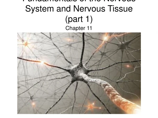
PART 1 Fundamentals of theNervous System andNervous Tissue
Nervous System • Master control and communication system • Has three overlapping functions • Sensory receptors monitor changes inside and outside the body • Change – a stimulus • Gathered information – sensory input
Nervous System • Processes and interprets sensory input • Makes decisions – integration • Dictates a response by activating effector organs • Response – motor output
Basic Divisions of the Nervous System • Central nervous system (CNS) • Brain and spinal cord • Integrating and command center
Basic Divisions of the Nervous System • Peripheral nervous system (PNS) • Outside the CNS • Consists of nerves extending from brain and spinal cord • Cranial nerves • Spinal nerves • Peripheral nerves link all regions of the body to the CNS • Ganglia are clusters of neuronal cell bodies
Basic Divisions of the Nervous System Figure 12.2
Sensory Input and Motor Output • Sensory (afferent) signals picked up by sensor receptors • Carried by nerve fibers of PNS to the CNS • Motor (efferent) signals are carried away from the CNS • Innervate muscles and glands
Sensory Input and Motor Output • Divided according to region they serve • Somatic body region • Visceral body region • Results in four main subdivisions • Somatic sensory • Visceral sensory • Somatic motor • Visceral motor
Types of Sensory and Motor Information Figure 12.3
Basic Divisions of the Nervous System • Somatic sensory • General somatic senses – receptors are widely spread • Touch • Pain • Vibration • Pressure • Temperature (receptors discussed in Chapter 14)
Basic Divisions of the Nervous System • Somatic sensory (continued) • Proprioceptive senses – detect stretch in tendons and muscle • Body sense – position and movement of body in space • Special somatic senses (Chapter 16) • Hearing • Balance • Vision • Smell
Basic Divisions of the Nervous System • Visceral sensory • General visceral senses – stretch, pain, temperature, nausea, and hunger • Widely felt in digestive and urinary tracts, and reproductive organs • Special visceral senses • Taste
Basic Divisions of the Nervous System • Somatic motor • General somatic motor – signals contraction of skeletal muscles • Under our voluntary control • Often called “voluntary nervous system” • Branchial motor • Typical skeletal muscle derived from somitomeres
Basic Divisions of the Nervous System • Visceral motor • Regulates the contraction of smooth and cardiac muscle • Makes up autonomic nervous system • Controls function of visceral organs • Often called “involuntary nervous system” • Autonomic nervous system (Chapter 15)
Nervous Tissue • Cells are densely packed and intertwined • Two main cell types • Neurons – transmit electrical signals • Support cells (neuroglial cells in CNS) • Nonexcitable • Surround and wrap neurons
The Neuron • The human body contains billions of neurons • Basic structural unit of the nervous system • Specialized cells conduct electrical impulses along the plasma membrane • Nerve impulse (action potential)
The Neuron • Other special characteristics • Longevity – can live and function for a lifetime • Do not divide – fetal neurons lose their ability to undergo mitosis; neural stem cells are an exception • High metabolic rate – require abundant oxygen and glucose • Neurons die after 5 minutes without oxygen
The Cell Body • Cell body (soma) • Perikaryon – around nucleus • Size of cell body varies from 5–140µm • Contains usual organelles plus other structures • Chromatophilic bodies (Nissl bodies) • Clusters of rough ER and free ribosomes • Stain darkly and renew membranes of the cell
The Cell Body • Neurofibrils – bundles of intermediate filaments • Form a network between chromatophilic bodies
The Cell Body • Most neuronal cell bodies • Located within the CNS • Protected by bones of the skull and vertebral column • Ganglia – clusters of cell bodies • Lie along nerves in the PNS
Structure of a Typical Large Neuron Figure 12.4
Neuron Processes • Dendrites • Extensively branching from the cell body • Transmit electrical signals toward the cell body • Chromatophilic bodies – only extend into the basal part of dendrites and to the base of the axon hillock • Function as receptive sites for receiving signals from other neurons
Neuron Processes • Axons • Neuron has only one • Impulse generator and conductor • Transmits impulses away from the cell body • Chromatophilic bodies are absent • No protein synthesis in axon
Neuron Processes • Axons (continued) • Neurofilaments, actin microfilaments, and microtubules • Provide strength along length of axon • Aid in the transport of substances to and from the cell body • Axonal transport
Neuron Processes • Axons • Branches along length are infrequent • Axon collaterals • Multiple branches at end of axon • Terminal branches (telodendria) • End in knobs called axon terminals(also called end bulbs or boutons)
Neuron Processes • Nerve impulse • Generated at the initial segment of the axon • Conducted along the axon • Releases neurotransmitters at axon terminals • Neurotransmitters – excite or inhibit neurons • Neuron receives and sends signals
Synapses • Site at which neurons communicate • Signals pass across synapse in one direction • Presynaptic neuron • Conducts signal toward a synapse • Postsynaptic neuron • Transmits electrical activity away from a synapse PLAY Synapse
Two Neurons Communicating at a Synapse Figure 12.6
Types of Synapses • Axodendritic • Between axon terminals of one neuron and dendrites of another • Most common type of synapse • Axosomatic • Between axons and neuronal cell bodies • Axoaxonic, dendrodendritic, and dendrosomatic • Uncommon types of synapses
Some Important Types of Synapses Figure 12.7
Synapses • Elaborate cell junctions • Axodendritic synapses – representative type • Synaptic vesicles on presynaptic side • Membrane-bound sacs containing neurotransmitters • Mitochondria abundant in axon terminals • Synaptic cleft • Separates the plasma membrane of the two neurons
Structure of a Synapses Figure 12.8a, b
Signals Carried by Neurons • Plasma membranes of neurons conduct electrical signals • Resting neuron – membrane is polarized • Inner, cytoplasmic side is negatively charged • Stimulation of the neuron depolarization
Signals Carried by Neurons Figure 12.9a, b
Signals Carried by Neurons • Strong stimulus applied to the axon triggers • Nerve impulse/action potential • Membrane becomes negative externally • Impulse travels the length of the axon • Membrane repolarizes itself
Signals Carried by Neurons Figure 12.9c–d
Synaptic Potentials • Excitatory synapses • Neurotransmitters alter the permeability of the postsynaptic membrane • Leads to an inflow of positive ions • Depolarizes the postsynaptic membrane • Drives the postsynaptic neuron toward impulse generation
Synaptic Potentials • Inhibitory synapses • The external surface of the postsynaptic membrane becomes more positive • Reduces the ability of the postsynaptic neuron to generate an action potential