Download
1 / 41
430 likes | 664 Views
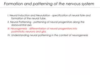
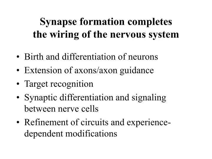
Synapse formation completes the wiring of the nervous system. Birth and differentiation of neurons Extension of axons/axon guidance Target recognition Synaptic differentiation and signaling between nerve cells Refinement of circuits and experience-dependent modifications.

E N D
Synapse formation completesthe wiring of the nervous system • Birth and differentiation of neurons • Extension of axons/axon guidance • Target recognition • Synaptic differentiation and signaling between nerve cells • Refinement of circuits and experience-dependent modifications
Synapse Formation in the Peripheral and Central Nervous System
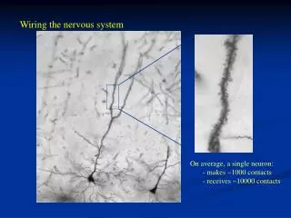
Synapses: the basic computation unitsin the brain • Human brain consists of 1011 neurons that form a network with 1014 connections • The number and specificity of synaptic connection needs to be precisely controlled • Changes of synaptic connections and synaptic strength are the basis of information processing and memory formation
Aberrant synaptic connectivityand synaptic function lead to disease states • Loss of synapses in Alzheimer’s disease • In epilepsy excessive synapse formation and synaptic misfunction are observed • Genes associated with mental retardation and schizophrenia have synaptic functions • Paralysis after spinal cord injuries
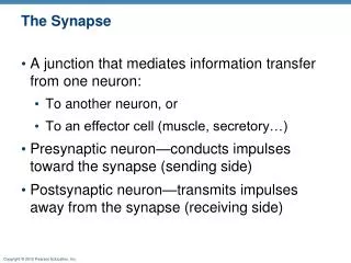
Central Synapses andNeuromuscular Junctions (NMJs) • Neuron-neuron and neuron-muscle synapses develop by similar mechanisms • NMJs are larger, more accessible and simpler than central synapses therefore the molecular mechanisms of synapse formation are best understood for the NMJ
Structure of the neuromuscular junction • Mature NMJs consist of three cell types • Motor nerve • Muscle cell • Schwann cells • All three cell types adopt a highly specialized organization that ensures proper synaptic function
Nerve terminal: - rich in synaptic vesicles - active zones - mitochondria - axon are rich in neurofilaments and contain only few vesicles
Muscle: - junctional folds opposing the active zones - specific cytoskeleton at synapse - strong concentration of ACh-R
Schwann Cells: - thin non-myelin processes that cover nerve terminal - myelin sheet around the remaining axon from exit site from the spinal cord to the NMJ
Basal Lamina: - present at synaptic and non-synaptic regions, but specific molecular composition at synapse (e.g.: acetylcholinesterase in cleft)
vesicles neurofilament ACh-receptors overlay
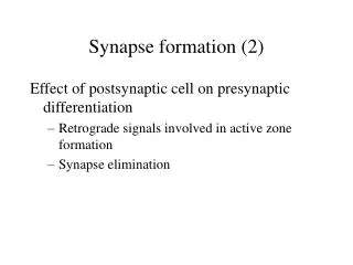
General Features of Synapse Formation 1) The pre- and post-synaptic cell organize each others organization (bi-directional signaling) 2) Synapses mature during development • widening of synaptic cleft, basal lamina • transition from multiple innervation to 1:1 3) Muscle and nerve contain components required for synaptogenesis (vesicles, transmitter, ACh-R) • “reorganization”
Stages of NMJ Development - growth cone approaches - non-specialized but functional contact - immature specializations - multiple innervation - elimination of additional axons, maturation
The basal lamina directs clustering of ACh-Rs Denervation and muscle elimination (but preservation of muscle satellite cells which will form new myotubes) In the absence of nerve, ACh-Rs cluster at the original synaptic site
Cultured muscle fiber Cultured muscle fiber + agrin Agrin • Component of the basal lamina • 400 kDa proteoglycan • Secreted from motor neuron and muscle • Neural form potently induces clustering of ACh-Rs in myotubes
Agrin signals through MuSK • agrin interacts with a MuSK/Masc on the muscle • MuSK is a receptor tyrosine kinase • MuSK activation leads to phosphorylation of • rapsyn and clustering of ACh-Rs
Mouse mutants confirm essential roles for agrin, MuSK, rapsyn Wild type Agrin mutant Rapsyn mutant MuSK mutant
Summary of mutant phenotypes • Agrin -/-: few ACh-R clusters, overshooting of axons • MuSK -/-: no ACh-R clusters, overshooting of axons • Rapsyn -/-: no ACh-R clusters, but higher receptor levels in synaptic area, only limited overshooting • Pre-synaptic defects in all mutants, due to the lack of retrograde signals from the muscle
A) Aggregation of existing receptors Agrin MuSK Rapsyn B) Local synthesis of receptors ???
Neuregulin (ARIA) • Acetylcholine receptor inducing activity • Expressed in motor neuron and in muscle • Binds and activates receptor tyrosine kinases on the muscle (erbB2, erbB3, erbB4) • Signals through MAP-kinase pathway • Leads to upregulation of ACh-R expression in sub-synaptic nuclei
Decrease in ACh-R in neuregulin (+/-) heterozygous mice Wild type Heterozygote MEPP (miniature excitatory potential)
Neural activity represses ACh-R synthesis in non-synaptic areas Paralysis Extra-synaptic ACh-R transcription increased Extra-synaptic ACh-R transcription decreased Denervation Electrical Stimulation Extra-synaptic ACh-R transcription increased Extra-synaptic ACh-R transcription decreased
Three neural signals for the induction of postsynaptic differentiation • Agrin: aggregation of receptors in the muscle membrane • Neuregulin: by upregulation of ACh-R expression in sub-synaptic nuclei • ACh/neural activity: downregulation of ACh-R expression in extra-synaptic nuclei
Components of the basal lamina can organize the nerve terminal Denervation + Muscle elimination Denervation Regeneration Regeneration
Laminin 11 affects presynaptic differentiation Wild type Lamininb2 mutant
Synaptic inactivity can lead to synapse elimination pre post pre post
Structure of excitatory synapses in the CNS Pre-synaptic terminal: Synaptic vesicles Pre-synaptic cytomatrix Active zone Synaptic cleft: 20 nm wide, filled with electron-dense material (proteins and carbohydrates) Post-synaptic compartment: Spine structure Dense submembrane scaffold Neurotransmitter receptors
Analogies of central synapses and NMJs • Overall structural similarities • Bi-directional signaling • Clustering of neurotransmitter receptors • Synaptic vesicles have similar components • Synapse elimination during development
Differences between central synapses and NMJs • No basal lamina • No junctional folds but dendritic spines • Multiple innervation is common • Difference in neurotransmitters: • Excitatory synapses use glutamate • Inhibitory synapses use GABA (g-aminobutyric acid) and glycine • different neurotransmitter receptors
Cytoplasmic scaffolding proteins mediate clustering of receptors in the CNS Gephryn clusters glycine receptors PSD95 clusters glutamate receptors • One neuron can receive excitatory and inhibitory inputs • through different synaptic connections • Transmitter in presynaptic vesicles is matched with the • postsynaptic receptors
Direct trans-synaptic interactions in the CNS neuroligin/ neurexin cadherins
Neuroligin can induce presynaptic differentiation in CNS neurons
Direct trans-synaptic interactions in the CNS neuroligin/ neurexin cadherins
Future directions/problems • Many factors that mediate synaptic differentiation in the CNS are not understood • Target specificity • Regeneration after injury is very low in CNS compared to PNS resulting in paralysis • Strategies to improve re-growth of axons and specific synapse formation