Download
1 / 18
200 likes | 407 Views
Medical Microbiology 480B: Bacteria: Structure and Function . Assigned reading: Murray, Chapters 1 and 2. Ren ée Tsolis, Ph.D. Office: 5519 Genome and Biomedical Sciences Facility rmtsolis@ucdavis.edu. The bacterial cell envelope. Extracellular structures: Flagella.

E N D
Medical Microbiology 480B: Bacteria: Structure and Function Assigned reading: Murray, Chapters 1 and 2 Renée Tsolis, Ph.D. Office: 5519 Genome and Biomedical Sciences Facility rmtsolis@ucdavis.edu
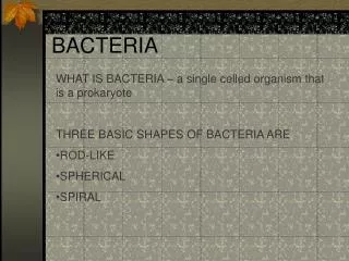
Extracellular structures: Flagella • Flagella are found on both gram-positive and gram-negative bacteria. • Flagella mediate swimming motility. The flagellum serves as a propeller that moves bacteria through a liquid medium. • Bacteria can sense concentration gradients and move toward nutrients or away from noxious stimuli. This is known as chemotaxis.
http://www.textbookofbacteriology.net/structure.html Extracellular structures: Flagella • Flagella may be polar (inserted at one end or pole of the bacterium) or peritrichous (inserted around the entire periphery of the cell). Polar flagella: Treponema denticola Peritrichous flagella: Salmonella typhimurium
Extracellular structures: Flagella Structure of the flagellum: The flagellar filament is composed of polymerized subunits called flagellin. Flagellin is also known as the H antigen. Since its antigenicity varies between bacterial species, it is used for typing (i.e. E. coli O157:H7). Flagellin is the ligand for Toll-like receptor 5 in innate immunity. http://www.textbookofbacteriology.net/structure.html
Extracellular structures: Pili Pili have been identified on both gram-positive and gram-negative bacteria. Morphologically, the filament of the pilus is thinner than that of the flagellum Pili have been shown to be involved in adhesion to cells and tissues. The scanning electron micrograph on the left shows enteropathogenic E. coli (EPEC)adhering to intestinal cells. E. coli expressing pili and flagella medschool.umaryland.edu/infeMSD/Images.htm
More about Pili A single bacterium may express more than one type of pilus (also called fimbriae). Pili have been shown to be involved in conjugative transfer of plasmids. This is medically significant, since plasmids often encode antibiotic resistance genes. Conjugative pili are also known as “sex pili”.
Extracellular structures: Capsule Many medically important bacteria produce a capsule. Capsules are generally made of polysaccharides, less frequently, protein may be present. Capsule often have an anti-phagocytic function, preventing uptake and killing by phagocytes. Some capsules are classified as antigens (Vi, K). Capsular polysaccharides are components of subunit vaccines (Typhoid, Pneumococcus). India Ink preparation of capsulated bacteria Electron micrograph of S. pyogenes Images: http://www.textbookofbacteriology.net/structure.html
Extracellular structures: Capsule vs. Slime Layer A true capsule is a discrete layer of substance surrounding the bacterium. Bacteria may also secrete polysaccharides that do not form a discrete structure; this is known as a slime layer or glycocalyx. Slime layers can provide a matrix for biofilm formation. In nature, biofilms generally contain mixed populations of microorganisms. Dental plaque biofilm containing Streptococcus mutans (www.denniskunkel.com) Biofilm on intravenous catheter containing Staphyococcus epidermidis and Pseudomonas aeruginosa (ASM Microbe Library)
Cell envelope: Gram + vs. Gram - Gram stain differentiates bacteria based on differences in the cell envelope. The thick peptidoglycan layer of Gram + bacteria traps the crystal violet. Peptidoglycan is also known as murein. The peptidoglycan layer in Gram + bacteria is 15-80 nm thick, while that of Gram - is only 10 nm. The cell wall forms a rigid structure that protects the cell from osmotic lysis. Gram + Gram - Top row: electron micrographs of the cell envelope; Bottom: a simplified scheme. Image: http://pathmicro.med.sc.edu/fox/cell_envelope.htm
Gram + cell envelope Teichoic acids and teichuronic acids are important constituents of the gram + cell wall. They are not present in gram-negative bacteria. These are polymers containing ribitol or glycerol joined through phosphodiester linkages and carrying amino acid or sugar residues. Teichoic acids bind Mg++ and are thought to supply it to the bacterium. Teichuronic acids are synthesized when phosphate is limiting and contain sugar acids in place of phosphoric acids. Staphylococcus aureus peptidoglycan. Reproduced from Jawetz, p. 23
Gram-negative cell envelope • Differences to Gram + • Outer membrane: contains lipoprotein, lipopolysaccharide (LPS) and proteins • Peptidoglycan: just 1 layer thick (compared to ~40 layers for gram+) • Periplasm: contains enzymes and transport proteins
Gram-negative cell envelope: LPS LPS is also known as endotoxin. It is the mediator of endotoxic shock during systemic infections. The O-antigen is a variable diagnostic marker and is used for serology (i.e. E. coliO157:H7). Lipid A is the toxic portion of LPS. It mediates toxicity via interaction with Toll-like receptor 4 (TLR4) of the innate immune system.
Cytoplasmic membrane • Present in both Gram + and Gram - • Contains phospholipids (40%) and proteins (60%) • Functions: • Nutrient uptake • Serves as a selective permeability barrier • Electron transport systems and ATP generation • Secretion of macromolecules • Chemotaxis • Coordination of DNA replication with cell division (septum formation)
Endospore formation • Some gram-positive bacteria can form endospores • Bacillus anthracis, Clostridium spp. are examples • Spore formation allows bacteria to survive unfavorable conditions (heat, dessication, radiation, chemicals). • Spores can survive for years in the environment • In bacteria: 1 bacterium gives rise to a single spore Pictures of endospores http://pathmicro.med.sc.edu/fox/cell_envelope.htm
Why is this relevant for the practice of medicine? • Peptidoglycan biosynthesis pathway is targeted by many antibiotics and gram stain will influence choice of therapy • Cell surface components are often antigenic and used in classification of bacteria • Cell surface components are often important pathogenicity determinants • Peptidoglycan, Lipid A, Lipoteichoic acid and flagellin are recognized by innate immunity and can induce inflammatory reactions
Important take-home points • Components of the cell envelope • Differences between Gram-positive and Gram-negative cell envelope • What is endotoxin and which bacteria make it? • What are the biological functions and properties of pili, flagella and capsules? • Which bacteria can form endospores and what is their function?