Download
1 / 38
420 likes | 894 Views
The Vertebral Column. Yes, this is a Predator Ewok…. The Vertebral Column. General Characteristics. Supports weight of trunk and distributes weight to lower limbs Surrounds and protects spinal cord Flexible curved structure composed of 26 irregular bones (vertebrae)

E N D
The Vertebral Column Yes, this is a Predator Ewok….
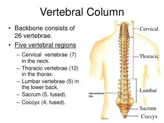
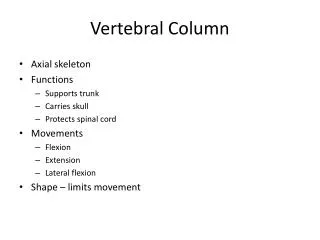
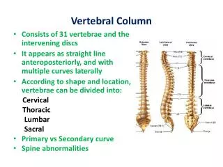
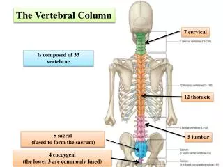
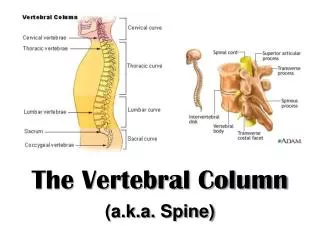
The Vertebral Column General Characteristics • Supports weight of trunk and distributes weight to lower limbs • Surrounds and protects spinal cord • Flexible curved structure composed of 26 irregular bones (vertebrae) • Cervical vertebrae (7)—vertebrae of the neck • Thoracic vertebrae (12)—vertebrae of the thoracic cage • Lumbar vertebrae (5)—vertebra of the lower back • Sacrum—bone inferior to the lumbar vertebrae • Coccyx—end of vertebral column
Vertebral Column: Curvatures • Increase the resilience and flexibility of the spine • Two concave curvatures • Cervical and lumbar • Two convex curvatures • Thoracic and sacral
Abnormal Spine Curvatures Scoliosis Kyphosis Lordosis
Ligaments • Anterior and posterior longitudinal ligaments • From neck to sacrum • Ligamentum flavum • Connects adjacent vertebrae
Intervertebral Discs • Cushionlike pad between vertebrae; acts like a shock absorber • Composed of two parts • Nucleus pulposus • Inner gelatinous nucleus that gives the disc its elasticity and compressibility • Anulus fibrosus • Outer collar composed of collagen and fibrocartilage • Limits the expansion of the nucleus pulposus when the spine is compressed • Withstands twisting forces on the spine and resists tension on spine
Intervertebral disc Supraspinous ligament Anterior longitudinal ligament Transverse process Sectioned spinous process Intervertebral foramen Posterior longitudinal ligament Ligamentum flavum Interspinous ligament Anulus fibrosus Nucleus pulposus Inferior articular process Sectioned body of vertebra Median section of three vertebrae, illustrating the composition of the discs and the ligaments Figure 7.17a
Vertebral spinous process (posterior aspect of vertebra) Spinal cord Spinal nerve root Transverse process Herniated portion of disc Anulus fibrosus of disc Nucleus pulposus of disc (c) Superior view of a herniated intervertebral disc Figure 7.17c
General Structure of Vertebrae • Body or centrum • Anterior weight-bearing region • Vertebral arch • Composed of pedicles and laminae that, along with the body, enclose vertebral foramen • Vertebral foramina • Together make up vertebral canal for spinal cord • Intervertebral foramina • Lateral openings between adjacent vertebrae for spinal nerves
General Structure of Vertebrae • Seven processes per vertebra: • Spinous process—projects posteriorly • Transverse processes (2)—project laterally • Superior articular processes (2)—protrude superiorly inferiorly • Inferior articular processes (2)—protrude inferiorly
Posterior Vertebral arch Lamina Spinous process Transverse process Superior articular process and facet Vertebral foramen Pedicle Body (centrum) Anterior Figure 7.18
CHECK POINT!!! What are the 5 major regions of the vertebral column? How many curvatures of the spine are there? And what are they? Which structure of the spine acts as a shock absorber?
Cervical Vertebrae • C1 to C7:smallest, lightest vertebrae • C1 (atlas) and C2 (axis) have unique features • C3 to C7 share the followingfeatures • Oval body • Spinous processes are bifid (except C7) • Large, triangular vertebral foramen • Transverse foramen in each transverse process
Cervical Vertebrae Atlas (C1) • No body or spinous process • No intervertebral disk between C1 and C2 • Consists of anterior and posterior arches, and two lateral masses • Superior surfaces of lateral masses articulate with the occipital condyles • Allow you to nod your head “yes”
Posterior Posterior C1 Posterior tubercle Posterior tubercle Posterior arch Inferior articular facet Posterior arch Lateral masses Transverse process Lateral masses Transverse foramen Superior articular facet Transverse foramen Anterior arch Anterior tubercle Anterior arch Facet for dens Anterior tubercle (a) Superior view of atlas (C1) (b) Inferior view of atlas (C1) Figure 7.19a-b
Dens projects superiorly into the anterior arch of the atlas • Dens is a pivot for the rotation of the atlas • Allows you to rotate head from side to side to say “no” Axis (C2)
Posterior C2 Spinous process Lamina Inferior articular process Pedicle Superior articular facet Transverse process Dens Body (c) Superior view of axis (C2) Figure 7.19c
Dens of axis Transverse ligament of atlas C1 (atlas) C2 (axis) C3 Inferior articular process Bifid spinous process Transverse processes C7 (vertebra prominens) (a) Cervical vertebrae Figure 7.20a
Thoracic Vertebrae • T1 to T12 • All articulate with ribs at facets and demifacets • Heart-shaped body • The vertebral foramen is circular • Long spinous process • Location of articular facets allows rotation of this area of spine
Superior articular process Transverse process Transverse costal facet (for tubercle of rib) Intervertebral disc Body Inferior costal facet (for head of rib) Spinous process Inferior articular process (b) Thoracic vertebrae Figure 7.20b
Lumbar Vertebrae • L1 to L5 • Short, thick pedicles and laminae • Flat hatchet-shaped spinous processes • Vertebral foramen is triangular • Orientation of articular facets locks lumbar vertebrae together so as to prevent rotation
Superior articular process Body Transverse process Intervertebral disc Inferior articular process Spinous process (c) Lumbar vertebrae Figure 7.20c
Sacrum 5 fused vertebrae (S1–S5) Forms posterior wall of pelvis Articulates with L5 superiorly, and with auricular surfaces of the hip bones laterally Coccyx Tailbone 3–5 fused vertebrae Articulates superiorly with sacrum Sacrum and Coccyx
Sacral promontory Ala Body of first sacral vertebra Transverse ridges (sites of vertebral fusion) Anterior sacral foramina Apex Coccyx (a) Anterior view Figure 7.21a
Facet of superior articular process Sacral canal Body Ala Auricular surface Median sacral crest Lateral sacral crest Posterior sacral foramina Sacral hiatus Coccyx (b) Posterior view Figure 7.21b
CHECK POINT!!! What are the 1st two cervical vertebra called and what makes them different? How can you tell the difference between a lumbar vertebrae and a thoracic vertebrae?
Thoracic Cage • Composed of • Thoracic vertebrae • Sternum • Ribs and their costal cartilages • Functions • Protects vital organs of thoracic cavity • Supports shoulder girdle and upper limbs • Provides attachment sites for many muscles, including intercostal muscles used during breathing
Sternum (Breastbone) • Three fused bones • Manubrium • Articulates with clavicles and ribs 1 and 2 • Body • Articulates with costal cartilages of ribs 2 through 7 • Xiphoid process • Site of muscle attachment • Not ossified until ~ age 40
Ribs and Their Attachments • 12 pairs • All attach posteriorly to thoracic vertebrae • Pairs 1 through 7 • True (vertebrosternal) ribs • Attach directly to the sternum by individual costal cartilages
Ribs and Their Attachments • Pairs 8 through12 • False ribs • Pairs 8–10 also called vertebrochondral ribs • Attach indirectly to sternum by joining costal cartilage of rib above • Pairs 11–12 also called vertebral (floating) ribs • No attachment to sternum
Jugular notch Clavicular notch Manubrium Sternal angle Body Sternum True ribs (1–7) Xiphisternal joint Xiphoid process False ribs (8–12) Intercostal spaces Costal cartilage Costal margin L1 Vertebra Floating ribs (11, 12) (a) Skeleton of the thoracic cage, anterior view Figure 7.22a
Structure of a Typical Rib • Main parts: • Head • Articulates posteriorly with facets (demifacets) on bodies of two adjacent vertebrae • Neck • Tubercle • Articulates posteriorly with transverse costal facet of same-numbered thoracic vertebra • Shaft
Transverse costal facet (for tubercle of rib) Superior costal facet (for head of rib) Angle of rib Body of vertebra Head of rib Intervertebral disc Neck of rib Tubercle of rib Shaft Sternum Cross- section of rib Costal groove Costal cartilage (a) Vertebral and sternal articulations of atypical true rib Figure 7.23a
Articular facet on tubercle of rib Spinous process Shaft Transverse costal facet (for tubercle of rib) Ligaments Neck of rib Body of thoracic vertebra Head of rib Superior costal facet (for head of rib) (b) Superior view of the articulation between arib and a thoracic vertebra Figure 7.23b
CHECK POINT!!! How does a true rib differ from a false rib? Besides the ribs and sternum, there is a 3rd group of bones that makes up the thoracic cage. What is it? What are floating ribs?