Download
1 / 39
861 likes | 2.3k Views
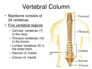
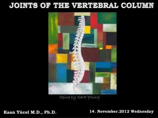
JOINTS OF THE VERTEBRAL COLUMN . Spine by Sara Young. 14. January.2014 Tuesday. Kaan Yücel M.D., Ph.D . JOINTS OF THE VERTEBRAL COLUMN. Symphyses between vertebral bodies n=2 1 above , 1 below Synovial joins between articular processes n=4 2 above , 2 below.

E N D
JOINTS OF THE VERTEBRAL COLUMN Spineby Sara Young 14. January.2014 Tuesday Kaan Yücel M.D., Ph.D.
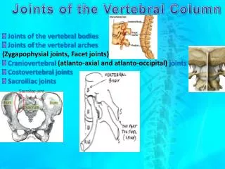
JOINTS OF THE VERTEBRAL COLUMN Symphysesbetweenvertebralbodiesn=2 1 above, 1 below Synovialjoinsbetweenarticularprocessesn=4 2 above, 2 below a total of 6 jointsbetweentwovertebrae 1) Craniovertebral(atlanto-axial and atlanto-occipital) joints 2) Costovertebraljoints 3) Sacroiliac joints
JOINTS OF THE VERTEBRAL BODIES Jointtype: symphyses (secondary cartilaginous joints) designed for weight-bearing and strength A layer of hyalinecartilageon eachvertebral body An intervertebraldiscbetweentheselayers an outer anulusfibrosussurrounds a central nucleus pulposus.
Anulusfibrosus an outer ring of collagen surrounding a wider zone of fibrocartilage arranged in a lamellar configuration.
Nucleuspulposus L. pulpa, fleshy core of the intervertebral disc fills the center of the intervertebral disc gelatinous absorbs compression forces between vertebrae. semifluid natureresponsible for much of the flexibility & resilience of the intervertebral disc and of the vertebral column as a whole
INTERVERTEBRAL DISCS Providestrong attachments between the vertebral bodies Unitevertebralbodiesinto a continuous semirigid column Formthe inferior ½ of anterior border of the intervertebral foramen. 20-25% of the length (height) of the vertebral column.
INTERVERTEBRAL DISCS Diagram of sagittal section of vertebral body and disc showing relationship of endplate and longitudinal ligament to the disc and the vertebrae. 1, vertebral body; 2, annulus fibrosus; 3, nucleus pulposus; 4, endplate; 5, spinal nerve root
INTERVERTEBRAL DISCS No intervertebral disc between C1 and C2 vertebrae Mostinferior functional disc between L5 and S1 vertebrae Anterior arch of the atlas (C1) Dens (odontoid peg around which atlas rotates) of axis (C2) Posterior arch of the atlas (C1) Soft palate (roof of the mouth) Root of the tongue Transverse process Intervertebral disc Inferior articular process Superior articular process Zygapophyseal(facet) joint Spinousprocess of C7 2nd-7th: The bodies of 2nd to 7th cervical vertebrae
INTERVERTEBRAL DISCS Thicknessof the discs vertebral column descends The range (amount) of movement relativethicknessto body greatest @ cervical &lumbar regions, movements of vertebral column greatestthickness most uniform in the thoracic region L4 L5
FUNCTIONS OF INTERVERTEBRAL DISCS Thankstothesemifluidnature Onevertebrarockforwardorbackward on anotherduringflexion & extension
INTERVERTEBRAL DISCS BY AGING • fibrocartilage. • Watercontent of thenucleuspulposus • Collagen fibers of the anulus degenerate. • Thin & lesselasticdiscs • Nucleus & anulus not distinguishable
JOINTS OF THE VERTEBRAL ARCHES Zygapophysial joints, FACET JOINTS plane synovial joints between superior &inferior articular processes of adjacent vertebrae. @ cervicalregionarticularcapsuleespeciallythin wide range of movement
JOINTS OF THE VERTEBRAL ARCHES Zygapophysial joints, FACET JOINTS permit gliding movements between articular processes shape &disposition of the articular surfaces determine the types of movement possible. Accessory ligaments unite the laminae, transverse processes, and spinous processes and help stabilize the joints.
UNCOVERTEBRAL (LUSCHKA’S) JOINTS Uncinateprocess elevations@ lateralmargins of theuppersurface Articulationwith the body of the vertebra above commonly between unciof the bodies of C3 or 4-C6 or 7 vertebrae @ the lateral &posterolateral margins of the intervertebral discs synovial joints ordegenerative spaces (clefts) in the discs occupied by extracellular fluid
LIGAMENTS OF THE VERTEBRAL COLUMN Joints between vertebrae reinforced &supported by numerous ligaments pass between vertebral bodies interconnect components of vertebral arches.
LIGAMENTS OF THE VERTEBRAL COLUMN Anterior & posterior longitudinal ligaments Ligamentaflava Supraspinousligament & ligamentumnuchae Interspinous ligaments betweentwolaminae
Posteriorlongitudinal ligament Anteriorlongitudinal ligament Tectorialmembrane Posteriorlongitudinalligamentconnectingaxistobase of theskull Frombase of the skull to anterior surface of sacrum Along its length attached to vertebral bodies and intervertebral discs
Ligamentaflava/Ligamentumflavum Between posterior surface of the lamina on the vertebra below anterior surface of the lamina of the vertebra above resist separation of the laminae in flexion. assist in extension back to the anatomical position.
Supraspinous ligament connects tips of thespinous processes from vertebra C7 to the sacrum From vertebra C7 to the skullbecomes structurally distinct ligamentumnuchae
Ligamentumnuchae • triangular, sheet-like structure in the median sagittal plane • Externaloccipitalprotuberancetomagnum • tip of spinousprocess of C7 • deep side attached to • posterior tubercle of vertebra C1 & spinousprocesses of other cervical vertebrae.
Ligamentumnuchae supports the head. resists flexion . facilitates returning the head to the anatomical position. provide attachment for adjacent musclesbroad lateral surfaces & posterior edge
Interspinousligaments between adjacent vertebral spinousprocesses • from base to apex of each spinous process • blend with • supraspinous ligament posteriorly • ligamentaflavaanteriorly • on each side
CRANIOVERTEBRAL JOINTS atlanto-occipital joints between atlas (C1) & occipital (condyle) bone atlanto-axialjoints between atlas (C1) & axis (C2) Synovialjointswithnointervertebraldiscs a wider range of movement than in the rest of the vertebral column.
ATLANTO-OCCIPITAL JOINTS Superiorarticularsurfaces of lateralmassesOccipitalcondyles nodding of the head, “yes” movement also sideways tilting of the head. Mainmovement flexion, with a little lateral flexion and rotation.
LIGAMENTS OF ATLANTO-OCCIPITAL JOINTS Anterior atlanto-occipital membrane(continuation of anteriorlong.lig.) connects anterior arch of the atlas to anterior margin of the foramen magnum Posterior atlanto-occipital membrane(similar to the ligamentumflavum) connects the posterior arch of the atlas to the posterior margin of the foramen magnum. help prevent excessive movement of the atlanto-occipital joints
ATLANTO-AXIAL JOINTS p Right & leftlateralatlantoaxialjoints betweeninf. facets of lateralmasses of C1 & superiorfacets of C2 Medianatlantoaxialjoint betweendens of axis& anteriorarch of atlas lane IVOT
MOVEMENTS OF ATLANTO-AXIAL JOINTS Cranium & atlas rotate on axis as a unit. Duringrotation of thehead Dens/pivot held in a collar anteriorlyanteriorarch of atlas posteriorlytransverseligament of atlas betweentubercles on medialsides of lateralmasses of atlas Headturns from sidetoside, disapproval (“no” movement).
LIGAMENTS OF ATLANTO-AXIAL JOINTS Superior and inferior longitudinal bands Apical ligament Alar ligaments Cruciate ligament of the atlas Tectorial membrane (Membranatectoria) .
COSTOVERTEBRAL JOINTS • A typical rib articulates with: • bodiesof adjacent vertebraejoint with the head of the rib • transverse process of its related vertebra costotransverse joint Necksrotatearoundtheirlongitudinalaxismainly in upperribs Ribsascenddescdendrelativetothespinemainly in lowerribs essential for altering the volume of the thoracic cavity during breathing
Joint with head of rib Head of therib Twofacetsface of articulation 1- withsuperiorfacet of itsownvertebra 2- withinferiorfacet of thevertebraabove divided into two synovial compartments by an intra-articular ligament
Costotransversejoints costotransverse ligamentmedialto the joint lateral costotransverse ligamentlateralto the joint attaches the tip of the transverse process to nonarticularpart of the tubercle of the rib. superior costotransverseligament attaches the superior surface of the neck of the rib to the transverse process of the vertebra above. Slight gliding movements
MOVEMENTS OF THE • VERTEBRAL COLUMN Rangeof movement according to the region and the individual Mobilityprimarily from compressibility & elasticity of the intervertebral discs Normalrange of movement reduced by 50% or more as a result of aging Movements by the vertebral column Flexion Extension Lateral flexion Rotation Circumduction
MOVEMENTS OF THE • VERTEBRAL COLUMN Movements in a specific region (cervical, thoracic, and lumbar) determined by shape &orientation of joint surfaces on the articular processes & on the vertebral bodies
MOVEMENTS OF THE • VERTEBRAL COLUMN • Rangeof movement limitedby • Thickness, elasticity, and compressibility of the IV discs • Shape &orientation of the zygapophysialjoints • Tension of the joint capsules of the zygapophysialjoints • Resistance of the back muscles and ligaments • Attachment to the thoracic (rib) cage • Bulk of surrounding tissue
DISC HERNIA & BACK PAIN A tear within the anulus fibrosus Material of the nucleus pulposus can track This material tracks into the vertebral canal or into the intervertebral foramen Pressure on neuralstructures commoncause of backpain
DISC HERNIA & BACK PAIN The use of diagnostic imaging in sports medicineMed J Aust 2005; 183: 482-486. Clinical Evaluation and Treatment Options for Herniated Lumbar Disc. Am Fam Physician. 1999;59:575-582. Degeneration and regeneration of the intervertebral disc: lessons from development. Dis Model Mech. 2011;4:31-41. Quantitative MRI as a diagnostic tool of intervertebral disc matrix composition and integrity.EurSpine J. 2008;17 Suppl4:432-440. Anatomy and pathophysiology of intervertebral disc disease. Techniques in Regional Anesthesia and Pain Management. Volume 13, 2009, Pages 67–75.
DISCECTOMY/LAMINECTOMY A prolapsed intervertebral disc may impinge upon meningeal sac spinal cord most commonly the nerve root producing symptoms attributable to that level. . PHYSIOTHERAPY OR INTERVENTION BY A NERUOSURGEON
DISCECTOMY/LAMINECTOMY Levelof the disc protrusion identified before surgery. MRI scanning and on-table fluoroscopy to prevent operating on the wrong level. In some instances removal of the lamina will increase the potential space and may relieve symptoms.