Download
1 / 41
450 likes | 792 Views
The Microbial World and You. Chapter 1 TFC. Microbiology The study of microorganisms. Microorganisms living things too small to be seen with the unaided eye Microorganisms= Microbes. Microbes in Our Lives. A few are pathogenic (disease-causing) Decompose organic waste

E N D
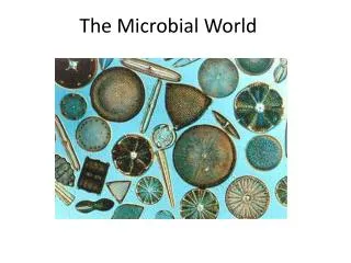
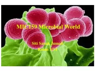
The Microbial World and You Chapter 1 TFC
MicrobiologyThe study of microorganisms • Microorganisms living things too small to be seen with the unaided eye • Microorganisms= Microbes
Microbes in Our Lives A few are pathogenic (disease-causing) Decompose organic waste Are producers in the ecosystem by photosynthesis Produce industrial chemicals such as ethanol and acetone Produce fermented foods such as vinegar, cheese, and bread Produce products used in manufacturing (e.g., cellulase) and treatment (e.g., insulin)
Designer Jeans: Made by Microbes? Stone-washing: Trichoderma Cotton: Gluconacetobacter Debleaching: Mushroom peroxidase Indigo: E. coli Plastic: Bacterial polyhydroxyalkanoate Applications of Microbiology, p. 3
Microbes in Our Lives Knowledge of microorganisms Allows humans to Prevent food spoilage Prevent disease occurrence Led to aseptic techniques to prevent contamination in medicine and in microbiology laboratories
Naming and Classifying Microorganisms Linnaeus established the system of scientific nomenclature Each organism has two names: the genus and specific epithet
Scientific Names Are italicized or underlined. The genus is capitalized, and the specific epithet is lowercase. Are “Latinized” and used worldwide. May be descriptive or honor a scientist.
Escherichia coli Honors the discoverer, Theodor Escherich Describes the bacterium’s habitat—the large intestine, or colon
Staphylococcus aureus Describes the clustered (staphylo-) spherical (cocci) cells Describes the gold-colored (aureus) colonies
Scientific Names After the first use, scientific names may be abbreviated with the first letter of the genus and the specific epithet: Escherichia coli and Staphylococcus aureus are found in the human body. E. coli is found in the large intestine, and S. aureus is on skin.
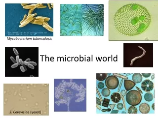
Types of Microorganisms Bacteria Archaea Fungi Protozoa Algae Viruses Multicellular animal parasites
Types of Microorganisms Figure 1.1
Bacteria Prokaryotes Peptidoglycan cell walls Binary fission For energy, use organic chemicals, inorganic chemicals, or photosynthesis Figure 1.1a
Archaea Prokaryotic Lack peptidoglycan Live in extreme environments Include Methanogens Extreme halophiles Extreme thermophiles Figure 4.5b
Fungi Eukaryotes Chitin cell walls Use organic chemicals for energy Molds and mushrooms are multicellular, consisting of masses of mycelia, which are composed of filaments called hyphae Yeasts are unicellular Figure 1.1b
Protozoa Eukaryotes Absorb or ingest organic chemicals May be motile via pseudopods, cilia, or flagella Figure 1.1c
Algae Eukaryotes Cellulose cell walls Use photosynthesis for energy Produce molecular oxygen and organic compounds Figure 1.1d
Viruses Acellular Consist of DNA or RNA core Core is surrounded by a protein coat Coat may be enclosed in a lipid envelope Viruses are replicated only when they are in a living host cell Figure 1.1e
Multicellular Animal Parasites Eukaryotes Multicellular animals Parasitic flatworms and roundworms are called helminths. Microscopic stages in life cycles. Figure 12.29
Classification of Microorganisms Three domains Bacteria Archaea Eukarya Protists Fungi Plants Animals
History of Microbiology • Microbes discovered >300yrs • Known to man during the mid 1800s • Period of progress began & continues to the present
Anton van Leeuwenhoek • 1674 made a simple microscope observed live specimens • Could magnify images up to 200x • Observed 50,000 different specimens, reported findings to the Royal Society of London
Spontaneous Generation • The formation of living things from inanimate objects • Was thought to be the origin of organisms • Disproved by Redi, Spallanzani, Pasteur
English Clergyman Needham (1774) • Proponent of spontaneous generation • Showed that boiling of meat broth had no effect on appearance of microbes, • Microbes developed spontaneously
Spontaneous Generation • Controversy continued for 100yrs • 1859 French Academy of Science competition to prove or disprove this theory
The Golden Age of Microbiology 1857–1914 Beginning with Pasteur’s work, discoveries included the relationship between microbes and disease, immunity, and antimicrobial drugs
Fermentation and Pasteurization Pasteur showed that microbes are responsible for fermentation Fermentationis the conversion of sugar to alcohol to make beer and wine Microbial growth is also responsible for spoilage of food Bacteria that use alcohol and produce acetic acid spoil wine by turning it to vinegar (acetic acid)
Fermentation and Pasteurization Pasteur demonstrated that these spoilage bacteria could be killed by heat that was not hot enough to evaporate the alcohol in wine Pasteurization is the application of a high heat for a short time Figure 1.4
Germ Theory of Disease • SG theory disproved led to rapid development of microbiology • Led to the study of infectious diseases
The Germ Theory of Disease 1835: Agostino Bassi showed that a silkworm disease was caused by a fungus 1865: Pasteur believed that another silkworm disease was caused by a protozoan 1840s: Ignaz Semmelweis advocated hand washing to prevent transmission of puerperal fever from one OB patient to another
The Germ Theory of Disease 1860s: Applying Pasteur’s work showing that microbes are in the air, can spoil food, and cause animal diseases, Joseph Lister used a chemical disinfectant to prevent surgical wound infections
The Germ Theory of Disease 1876: Robert Koch proved that a bacterium causes anthrax and provided the experimental steps, Koch’s postulates, to prove that a specific microbe causes a specific disease Figure 1.4
German Physician Koch (1876) • Proved that microorganisms caused diseases • Only specific microorganisms caused specific diseases • Studied anthrax affects cattle & humans
Immunity /Vaccination • Edward Jenner ( 1796) Smallpox immunity / Vaccine • Pasteur ( 1800s) vaccines for anthrax, rabies attenuated organisms
The Birth of Modern Chemotherapy Treatment with chemicals is chemotherapy Chemotherapeutic agents used to treat infectious disease can be synthetic drugs or antibiotics Antibiotics are chemicals produced by bacteria and fungi that inhibit or kill other microbes
The First Synthetic Drugs Quinine from tree bark was long used to treat malaria Paul Erlich speculated about a “magic bullet” that could destroy a pathogen without harming the host 1910: Ehrlich developed a synthetic arsenic drug, salvarsan, to treat syphilis 1930s: Sulfonamides were synthesized
A Fortunate Accident—Antibiotics 1928: Alexander Fleming discovered the first antibiotic Fleming observed that Penicillium fungus made an antibiotic, penicillin, that killed S. aureus 1940s: Penicillin was tested clinically and mass produced Figure 1.5