Download
1 / 72
730 likes | 1.15k Views
The Microbial World And You Chapter 1. Biotech I . Microbes in Our Lives. Microorganisms are organisms that are too small to be seen with the unaided eye. Microorganisms :. Decompose organic waste Are producers in the ecosystem by photosynthesis (they make sugars that we use)

E N D
The Microbial World And You Chapter 1 Biotech I
Microbes in Our Lives • Microorganisms are organisms that are too small to be seen with the unaided eye.
Microorganisms: • Decompose organic waste • Are producers in the ecosystem by photosynthesis (they make sugars that we use) • Produce industrial chemicals such as ethyl alcohol and acetone • Produce fermented foods such as vinegar, cheese, and bread • Some are pathogenic (disease causing)
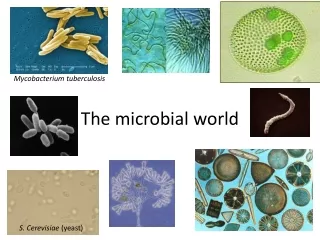
Microorganisms: Bacteria Fungi Algae Virus Protozoa Figure 1.1
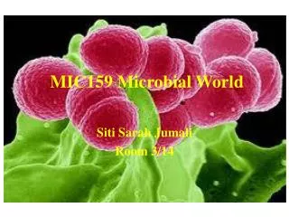
Bacteria • Prokaryotes • Peptidoglycan cell walls • Binary fission • For energy, use organic chemicals, inorganic chemicals, or photosynthesis Figure 1.1a
Archaea: • Prokaryotic • Lack peptidoglycan • Live in extreme environments • Include: • Methanogens • Extreme halophiles • Extreme thermophiles Figure 4.5b
Fungi • Eukaryotes • Chitin cell walls • Use organic chemicals for energy • Molds and mushrooms are multicellular • Yeasts are unicellular(single cell) Figure 1.1b
Protozoa • Eukaryotes • Absorb or ingest organic chemicals • May be motile via pseudopods, cilia, or flagella Figure 1.1c
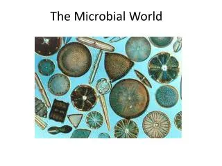
Algae • Eukaryotes • Cellulose cell walls • Use photosynthesis for energy • Produce molecular oxygen and organic compounds Figure 1.1d
Viruses • Acellular (ie. Without a cell) • Consist of DNA or RNA core • Core is surrounded by a protein coat • Coat may be enclosed in a lipid envelope • Viruses are replicated only when they are in a living host cell Figure 1.1e
Multicellular Animal Parasites • Eukaryote • Multicellular animals • Parasitic flatworms and round worms are called helminths. • Microscopic stages in life cycles. Figure 12.28
Classification of Organisms • Three domains • Bacteria • Archaea • Eukarya • Protists • Fungi • Plants • Animals
Microbes and Human Welfare • Microbial Ecology • Bacteria recycle carbon, nutrients, sulfur, and phosphorus that can be used by plants and animals.
Biological Insecticides • Microbes that are pathogenic to insects are alternatives to chemical pesticides to prevent insect damage to agricultural crops and disease transmission. • Bacillus thuringiensis infections are fatal in many insects but harmless to other animals including humans and to plants.
Modern Biotechnology and Genetic Engineering • The use of microbes to produce foods and chemicals, is centuries old. • Genetic engineering is a new technique for biotechnology. Through genetic engineering, bacteria and fungi can produce a variety of proteins including vaccines and enzymes. • Missing or defective genes in human cells can be replaced in gene therapy. • Genetically modified bacteria are used to protect crops from insects and freezing.
1700sCompeting Theories About Life • Abiogenesis: "spontaneous generation“ life develops from non-life • Biogenesis: life develops from life
Evidence Pro and Con • 1668: Francisco Redi filled six jars with decaying meat.
Evidence Pro and Con • 1745: John Needham put boiled nutrient broth into covered flasks.
Evidence Pro and Con • 1765: Lazzaro Spallanzani boiled nutrient solutions in flasks.
Evidence Pro and Con • 1861: Louis Pasteur demonstrated that microorganisms are present in the air.
The Theory of Biogenesis • Pasteur’s S-shaped flask kept microbes out but let air in. Figure 1.3
The Ubiquity of Microorganisms Ubiquity = existing everywhere Microorganisms live everywhere… in you, on you, in your food, in your bed, on the floor of your bathroom, on doorknobs, on EVERYTHING that’s not sterilized! For every one of your human cells, there are 10 bacterial cells! The bacteria that live in and on you naturally are your NORMAL FLORA
Units of Measurement • 1 µm = 10-6 m = 10-3 mm • 1 nm = 10-9 m = 10-6 mm • 1000 nm = 1 µm • 0.001 µm = 1 nm
Microscopy: The Instruments • A simple microscope has only one lens. Figure 1.2b
Microscopy: The Instruments • In a compound microscope the image from the objective lens is magnified again by the ocular lens. • Total magnification =objective lens ocular lens Figure 3.1b
Microscopy: The Instruments • Resolution is the ability of the lenses to distinguish two points. • A microscope with a resolving power of 0.4 nm can distinguish between two points ≥ 0.4 nm. • Shorter wavelengths of light provide greater resolution.
Microscopy: The Instruments • Refractive index is the light-bending ability of a medium. • The light may bend in air so much that it misses the small high-magnification lens. • Immersion oil is used to keep light from bending. Figure 3.3
Brightfield Illumination • We use in lab • Dark objects are visible against a bright background. • Light reflected off the specimen does not enter the objective lens. Figure 3.4a, b
Darkfield Illumination • Light objects are visible against a dark background. • Light reflected off the specimen enters the objective lens. Figure 3.4a, b
Phase-Contrast Microscopy • Accentuates diffraction of the light that passes through a specimen. Figure 3.4c
Differential Interference Contrast Microscopy • Accentuates diffraction of the light that passes through a specimen; uses twobeams of light. Figure 3.5
Fluorescence Microscopy • Uses Ultra Violet (UV) light. • Fluorescent substances absorb UV light and emit visible light. • Cells may be stained with fluorescent dyes (fluorochromes). Figure 3.6b
Confocal Microscopy • Uses fluorochromes and a laser light. • The laser illuminates each plane in a specimen to produce a 3-D image. Figure 3.7
Electron Microscopy • Uses electrons instead of light. • The shorter wavelength of electrons gives greater resolution.
Transmission Electron Microscopy (TEM) • Ultrathin sections of specimens. • Electrons pass through specimen, then an electromagnetic lens, to a screen or film. • Specimens may be stained with heavy metal salts. Figure 3.8a
Transmission Electron Microscopy (TEM) • 10,000-100,000; resolution 2.5 nm Figure 3.8a
Scanning Electron Microscopy (SEM) • An electron gun produces a beam of electrons that scans the surface of a whole specimen. • Secondary electrons emitted from the specimen produce the image. Figure 3.8b
Scanning Electron Microscopy (SEM) • 1000-10,000; resolution 20 nm Figure 3.8b
Scanning-Probe Microscopy • Scanning tunneling microscopy uses a metal probe to scan a specimen. • Resolution 1/100 of an atom. Figure 3.9a
Scanning-Probe Microscopy • Atomic force microscopy uses a metal and diamond probe inserted into the specimen. • Produces 3-D images. Figure 3.9b
Obtaining Pure cultures • A pure culture contains only one species or strain • A colony is a population of cells arising from a single cell or spore or from a group of attached cells • A colony is often called a colony-forming unit (CFU)
Streak Plate Figure 6.10a, b
Isolation Streak • Goal: to isolate single colonies of bacteria Correct Incorrect
1 4 2 3 loop
Average size of prokaryotes: 0.2 -1.0 µm 2 - 8 µm • Basic shapes:
Prokaryotic Arrangements • Pairs: diplococci, diplobacilli • Clusters: staphylococci • Chains: streptococci, streptobacilli
Some prokaryotes have unusual shapes • Star-shaped Stella • Square Haloarcula • Most bacteria are monomorphic (they only exist in one shape) • A few are pleomorphic (they can take different shapes) Figure 4.5