Download

1 / 41
460 likes | 849 Views
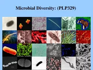
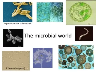
DIVERSITY OF THE MICROBIAL WORLD. Microbial World. A major biologic division separates the eukaryotes and prokaryotes

E N D
MicrobialWorld • A major biologic division separates the eukaryotes andprokaryotes • Cells from animals, plants, and fungi are eukaryotes (Greek for "true nucleus"), whereas bacteria and blue-green algae belong to the prokaryotes (Greek for "primitive nucleus"). www.themegallery.com CompanyLogo
Prokaryotes -Eukaryotes www.themegallery.com CompanyLogo
Prokaryotes -Eukaryotes www.themegallery.com CompanyLogo
Taxonomy andClassification • Classification, nomenclature, and identification are the three separate but interrelated areas of taxonomy • Classification can be defined as the arrangement of organisms into taxonomic groups (taxa) on the basis of similarities or relationships • Nomenclature is naming an organism by international rules according to its characteristics www.themegallery.com CompanyLogo
Taxonomy andClassification • Identification refers to the practical use ofa • classification scheme: • (1) to isolate and distinguish desirable organisms from undesirableones; • (2) to verify the authenticity or special properties of a culture; or, in a clinicalsetting, • (3) to isolate and identify the causative agent of adisease. • refers to the classification and grouping of organisms • based on genotypic (genetic) andphenotypic (observable) similarities anddifferences www.themegallery.com CompanyLogo
Taxonomy andClassification • Phenotypic • Classificationof Bacteria: • Microscopic morphology • Macroscopic morphology • Biotyping • Serotyping • Antibiogrampatterns • Phage typing • Genotypic Classificationof Bacteria: • Guanine plus cytosine ratio • DNA hybridization • Nucleic acidsequence analysis • Plasmidanalysis • Ribotyping • ChromosomalDNA fragmentanalysis www.themegallery.com CompanyLogo
NumericalTaxonomy • Also called computer taxonomy, phenetics, ortaxometrics • Numerical classification schemes use a large number (frequently 100 or more) of unweighted taxonomically useful characteristics • The computer clusters different strains at selected levels of overall similarity(usually • > 80% at the species level) on the basisof • the frequency with which they sharetraits. www.themegallery.com CompanyLogo
Taxonomy • According to a proposal by Woese, the world of living things is classified in the three domains bacteria, archaea, and eucarya. In this system, each domain is subdivided into kingdoms. • Bacteria: • heterotrophic eubacteria all human pathogen bacteria • photosynthetic cyanobacteria not pathogenic www.themegallery.com CompanyLogo
Taxonomy • Bacteria: • Classic bacteria reproduce asexually by binary transversefission • Chlamydiae obligate intracellularparasites • Rickettsiae obligate intracellular parasites, rod shaped to coccoid, that reproduce by binary transverse fission • Mycoplasmas bacteria without rigid cell walls www.themegallery.com CompanyLogo
Therankoftaxonomycanbeseeninthistable: Grading Example Kingdom Prokaryote Division Gracilicutes Class Scotobacteria Order Eubacteriales Family Enterobacteriaceae Genus Escherichia Species coli www.themegallery.com CompanyLogo
Nomenclature • provides naming assignments for each organism • family name is capitalized and hasan- • aceae ending (e.g.,Micrococcaceae) • genus name is capitalized and followed by the species name, begins with a lowercase letter; should be italicized in print but underlinedin the script (e.g., Staphylococcus aureus or Staphylococcus aureus) www.themegallery.com CompanyLogo
Nomenclature • using the first letter of the genus followed by a period and the species epithet (name) (e.g., S.aureus) • Species abbreviated sp. (singular) or spp. (plural) is used when the species is not specified • When referred to as a group, their names are neither capitalized nor underlined (e.g.,staphylococci) • The plural of genus is genera (e.g., Enterobacteriaceae family) www.themegallery.com CompanyLogo
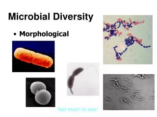
Morphology andStructure • Although bacteria are difficult to differentiate by size, they do have different shapes • Bacteria vary in size from 0.4 to 2 m occur in three basicshapes: • Cocci(spherical) • Bacilli(rod-shaped) • Spirochetes(helical) • Cocci: singly, pairs (diplococci), chains (streptococci), clusters(staphylococci) www.themegallery.com CompanyLogo
Morphology andStructure www.themegallery.com CompanyLogo
Morphology andStructure Bacilli: very short coccobacilli long filamentous rods, ends may be square or rounded • Bacilli with tapered, pointedends • fusiform • a species varies in size and shape within a pure culture pleomorphic • Bacilli may occur as single rods or in chains or may align themselves sideby side(palisading) www.themegallery.com CompanyLogo
Major Characteristics of Eukaryotesand Prokaryotes Eukaryote Prokaryote Algae, fungi,protozoa,plants, Bacteria animals >5 μm 0.5-3.0μm Characteristic Majorgroups Size(approximate) NuclearStructures Nucleus Classicmembrane No nuclearmembrane Chromosomes Strands of DNA Diploid genome Single, circular DNA Haploid genome Cytoplasmic Structures Mitochondria Present Absent Golgibodies Present Absent Endoplasmic reticulum Present Absent Ribosomes (sedimentation coefficient) 80S (60S+40S) 70S (50S+30S) www.themegallery.com CompanyLogo
Major Characteristics of Eukaryotesand Prokaryotes Eukaryote Prokaryote Containssterols Does not containsterols Characteristic Cytoplasmic membrane Cellwall Present for fungi; otherwise absent Is a complex structure containing protein, lipids, and peptidoglycans Reproduction Sexual andasexual Asexual (binary fission) Movement Complex flagellum, ifpresent Simple flagellum, ifpresent Respiration Viamitochondria Via cytoplasmicmembrane www.themegallery.com CompanyLogo
Eukaryotic CellStructure • The nucleus contains the cell'sgenome • The inner membrane is usually a simple sac, but the outermost membrane is continuous with the endoplasmicreticulum • The chromosomes of eukaryotic cells contain linear DNA macromolecules arranged as a double helix. • A structure often visible within thenucleus • is the nucleolus, an area rich in RNAthat • is the site of ribosomal RNAsynthesis. www.themegallery.com CompanyLogo
CytoplasmicStructures • The cytoplasm of eukaryotic cells is characterized by the presence of an endoplasmic reticulum, vacuoles, self- reproducing plastids, and an elaborate cytoskeleton composed of microtubules, microfilaments, and intermediatefilaments. • A variety of anaerobic or aerotolerant eukaryotic microorganisms (eg, Trichomonas vaginalis) lack mitochondria and contain instead a membrane-enclosed respiratory organelle called thehydrogenosome. www.themegallery.com CompanyLogo
CytoplasmicStructures • The cytoplasm is enclosed within a plasma membrane composed of protein and phospholipid, similar to the prokaryotic cellmembrane • Many eukaryotic microorganisms have organelles called flagella or cilia that move with a wave-like motion to propel the cell throughwater • Eukaryotic flagella emanate from the polar region of the cell, whereas cilia, whichare shorter than flagella, surround thecell www.themegallery.com CompanyLogo
Flagella • Flagella is accessory structure of bacteria, seen filamentous, is built from protein, only found at bacillus shape bacteria. Flagella is used in bacterial movement. • Depends on amount and catching to bacteria it is classified in: • Atricous :unflagella • Monotricous : single flagella on one ofpole • Lophotricous : more than 2 flagella on one ofpole • Amphitricous : more than 2 flagella on eitherpoles • Peritricous: many flagellasurrounding cellbody ofbacteria www.themegallery.com CompanyLogo
Prokaryotic CellStructure The nucleoid of bacterial cells has long been considered to consist of a single continuous circular molecule with a molecular weight of approximately 3 x 10⁹. It may thus be considered to be a single, haploid chromosome, approximately 1 mm long in the unfoldedstate www.themegallery.com CompanyLogo
CytoplasmicStructures • Prokaryotic cells lack autonomous plastids, such as mitochondria and chloroplasts; the electron transport enzymes are localized instead in the cytoplasmicmembrane • Microtubular structures, which are characteristics of eukaryotic cells, are generally absent inprokaryotes www.themegallery.com CompanyLogo
The CellEnvelope Bacteria are classified as gram-positive or gram-negative according to their response to the Gram staining procedure named for the histologist HansChristianGram: who developed this differential staining procedure in an attempt to stain bacteria in infected tissues. • The cells are first stained with crystal violet and iodine and then washed with acetone or alcohol. The latter step decolorizes gram- negative bacteria but not gram-positive bacteria. www.themegallery.com CompanyLogo
The CellEnvelope The difference between gram-positive and gram-negative bacteria has been shown to reside in the cell wall: Gram-positive cells can be decolorized with acetone or alcohol if the cell wall is removed after the staining step but before the washing step. Although the chemical composition of gram-positive and gram-negative walls is now fairly well known (see below), the reason gram-positive walls block the dye- extraction step is stillunclear. www.themegallery.com CompanyLogo
GramStaining www.themegallery.com CompanyLogo
Bacterial MembraneStructures Structure ChemicalConstituents Plasmamembrane Phospholipids, proteins, and enzymes involved in generation of energy, membrane potential, and transport Cell Wall Gram-positivebacteria Peptidoglycan Glycan chains of GlcNAc and MurNAc cross-linked by peptide bridge Teichoicacid Polyribitol phosphate or glycerol phosphate cross-linked topeptidoglycan Lipoteichoicacid Lipid-linked teichoicacid Gram-negativebacteria Peptidoglycan Thinner version of that found in gram-positivebacteria Periplasmicspace Enzymes involved in transport, degradation, and synthesis Outermembrane Phospholipids with saturated fattyacids www.themegallery.com CompanyLogo
Bacterial MembraneStructures Structure ChemicalConstituents Proteins Porins, lipoprotein, transportproteins LPS Lipid A, core polysaccharide, Oantigen Otherstructures Capsule Polysaccharides (disaccharides and trisaccharides) and polypeptides Pili Pilin,adhesins Flagellum Motor proteins,flagellin Proteins M protein of streptococci (as anexample) www.themegallery.com CompanyLogo
Pathogenesis of the BacterialInfections • The terms pathogenicity and virulence are not clearly defined in their relevance to microorganisms. They are sometimes even usedsynonymously. • It has been proposed that pathogenicity be used to characterize a particular species and that virulence be used to describe the sum of the disease-causing properties of a population (strain) of a pathogenic species www.themegallery.com CompanyLogo
Pathogenesis • Relatively little is known about the factors determining the pathogenicity and virulence of microorganisms, and most of what we do know concerns the disease- causing mechanisms ofbacteria. • Pathogenicity Capacity of a pathogen species to cause disease • Virulence Sum of thedisease-causing properties of a strain of a pathogenic species www.themegallery.com CompanyLogo
Incubation &Colonization • Incubation period Time between infection and manifestation of disease symptoms; this specific disease characteristic can be measured in hours, days, weeks, or even years • Colonization Presence of microorganisms on skin or mucosa; no penetration into tissues; typical of normal flora; pathogenic microorganisms occasionally also show colonizationbehavior www.themegallery.com CompanyLogo
Infection • Infection Invasion of a host organism by microorganisms, proliferation of the invading organisms, and hostreaction • Inapparent (or subclinical) infection Infection without outbreak of clinical symptoms • Infectious disease (or clinical infection) Infection with outbreak of clinical symptoms www.themegallery.com CompanyLogo
Infection • Probability of manifestation Frequencyof • clinical manifestation of an infection in disposed individuals (%) • Endogenous infection Infection arising from the colonizing flora • Exogenous infection Infection arising from invasion of host by microorganisms from sources external toit • Nosocomial infection Infection acquired during hospitalization (urinary tract infections,infections of the respiratory organs, wound infection, sepsis) www.themegallery.com CompanyLogo
Infection • Local infection Infection that remains restricted to the portal of entry and surroundingarea • Generalized infection Lymphogenous and/ or hematogenous spread of invading pathogen starting from the portal of entry; infection of organs to which pathogen shows a specific affinity (organotropism); three stages: incubation, generalization, organ manifestation www.themegallery.com CompanyLogo
Infection • Transitorybacteremia/viremia/parasitemia • Brief presence of microorganisms in the bloodstream • Superinfection: Occurrence of a second infection in the course of a firstinfection • Relapses: Series of infections by the samepathogen • Reinfection: Series of infections by different pathogens www.themegallery.com CompanyLogo
Pathogenesis of infectiousdisease There are five groups of potentialbacterial contributors to the pathogenesis of infectious diseases: Adhesins. They facilitate adhesion to specific target cells. Invasins. They are responsible for active invasion of the cells of themacroorganism. Impedins. These components disable host immune defenses in somecases. Aggressins. These substances include toxins and tissue-damagingenzymes. Modulins. Substances that induce excesscytokine production (i.e., lipopolysaccharides of Gram-negative bacteria, superantigens, mureinfragments). www.themegallery.com CompanyLogo
Invasion andSpread • Invasion. Bacteria may invade a host passively through microtraumata or macrotraumata in the skin or mucosa. On the other hand, bacteria that invade through intact mucosa first adhere to this anatomical barrier, then actively breachit. • Spread. Local tissue spread beginning at the portal of entry, helped along by tissue- damaging exoenzymes (hyaluronidase, collagenase, elastase, and other proteases). www.themegallery.com CompanyLogo
Spread Cell-to-cell spread. Bacteria translocated into the intracellular space by endocytosis cause actin to condense into filaments, which then array at one end of the bacterium and push up against the inner side of the cell membrane. This is followed by fusion with the membrane of the neighboring tissue cell, whereupon the bacterium enters the new cell (typical of Listeria andShigella). www.themegallery.com CompanyLogo
Virulence, Pathogenicity, Susceptibility,Disposition www.themegallery.com CompanyLogo
Alhamdulillah LOGO