Download
1 / 84
860 likes | 1.25k Views
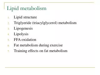
Lipid metabolism I. Biochemistry I Lecture 8 20 13 ( E.T. ). Major classes of lipids. triacylglycerols. prostanoids leukotriens. Energy nutrients. steroids. Derived lipids. phospholipids. glycerophospholipids. sphingofosfolipids.

E N D
Lipid metabolism I Biochemistry I Lecture 8 2013 (E.T.)
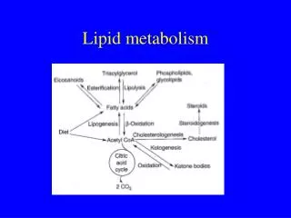
Major classes of lipids triacylglycerols prostanoids leukotriens Energynutrients steroids Derivedlipids phospholipids glycerophospholipids sphingofosfolipids Mainlystructuralcomponentsofmembranes 2
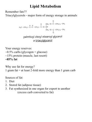
Metabolisms of lipids metabolism of TG a FA 100 g/day Source of energy metabolism of structural lipids 2 g/day Triacylaglycerols are the most effective form of energy deposition.
Triacylglycerols, fatty acids and esterified cholesterol are very hydrophobic they are not soluble in water unless they are emulsified or included in micelles in the presence of tensides.
Emulsificationoflipids in theintestine – formationofmicelles colipase Themaintensides are fattybileacids
Lipid adsorption in theintestine Formationofmixedmicellesfromproductsofdigestion Mixedmicellesare composed of fatty acids, mono/diacylglycerols, bile acids, phospholipids and fat-soluble vitamins Bile acids, phospholipids and fattyacidsfunction as tensides Intestinal lumenMucosal cell (enterocyte)
In the extracellular fluids hydrophobic lipids are transported in the form of lipoprotein particles
Lipoprotein particles transport triacylglycerols and cholesterol in body fluids Hydrophobic core Superficial layer (hydrophilic surface) monolayer
TG PL CH Proteiny Types of lipoproteins Chylomikron CM VLDL (very low density) HDL (high density) LDL (low density 11
Metabolism of triacylglycerols 1. Hydrolytic cleavage of ester bonds 2. Metabolism of fatty acids and glycerol 12
–C–O–CH O O O CH2–O–C– CH2–O–C– Lipases are enzymes that catalyse hydrolysis of ester bonds of triacylglycerols releasingfree fatty acids. Extracellular lipases Pancreatic lipasesecreted into the duodenum, Lipoprotein lipaseon the surface of the endothelium lining the capillaries Intracellular lipases Hormone-sensitive lipase of adipocytesmobilizing fat stores Lysosomal lipase
Transport of fatty acids in ECT FA are released mainly from TG in adipocytes by the action of hormon-sensitive lipase (hormonal regulation) or from lipoprotein particles FA in blood – carried by albumin (1 mmol/l, half-life 2 min) Transport of FA in cells • special membrane proteins facilitate the transport of FA across cytoplasmatic membrane • fatty acid binding proteins facilitate the intracellular transport • carnitine facilitates the transport across mitochondrial membrane 14
Degradation of lipids in the body Chylomicron, VLDL liver, muscle LP-lipase mitochondrie adipocytes ER FA Binding to albumin HS-lipase FA Binding to FABP -oxidation TG Binding to carnitin acetylCoA Hormone-sensitive lipase in adipocytes is an intracellular lipase that through hydrolysis of triacylglycerolsmobilizes the fat energy reserves. The activity of this lipase is controlled by hormones: Glucagon (at low blood glucose) and adrenaline/noradrenaline(in stress)
Degradation of fatty acids: β-oxidation Fatty acids serve as an energy sourcefor most of the cells (not for the nervous system and for red blood cells). The tissues gain fatty acids - either from lipoprotein particles after the triacylglycerols have been hydrolysedby lipoprotein lipase, - or as fatty acids mobilized by the action of hormones on the fat stores in adipose tissue and supplied bound onto albumin. Location: matrix of mitochondria
The utilization of fatty acids in the cells requires three stages of processing 1.Activation of FA by linking to coenzyme A 2.Transport of acyl CoA into themitochondrial matrix 3. β-Oxidation of acyl CoA in the mitochondrial matrix to acetyl CoA that enters the citrate cycle.
N H 2 Coenzyme A N N N N O O C H 3 O ~ P O P O C H C C H C C H O O 2 2 C C H C H H N O 2 2 C H O O H O 3 HS C H C H H N 2 2 β-Alanine Pantoic acid Cysteamine Pantothenic acid O O H O O P O 3´–phospho ADP 1. Activation of a fatty acid – synthesis of acyl coenzyme A Acyls can be attached to the sulfanyl group by means of a thioester bond.
The synthesis of the high-energy acyl-CoA thioester is catalysed by acyl-CoA synthetases R–COO– + CoA–SH R–CO–S-CoA Acyl-CoA synthetases are located on the outer mitochondrial membrane. There is a loss of energy equivalent to 2 molecules of ATP, because the reaction is made irreversible by the hydrolysis of inorganic diphosphate (AMP + ATP 2 ADP). AMP + 2 Pi ATP Synthesisof acyl-CoA
CH3 – CH2–CH–CH2–COO H3C N CH3 OH • Carnitine carries long-chain activated fatty acids into the mitochondrial matrix Acyl-CoA itself cannot cross the inner mitochondrial membrane; instead, acyl groups are transferred to carnitine, transported across the membrane asacylcarnitine, and transferred back to CoA within the mitochondrial matrix. Short-chain fatty acids (4 – 10 carbon atoms) do not require the carnitine shuttle, they can cross the inner mitochondrial membrane. Carnitine Trimethyl(2-hydroxy-3-carboxypropyl)ammonium
C H 3 H C N C H C H C H C O O H 3 2 2 O C H 3 C O The transfers of acyls from acyl-CoA to carnitine and from acylcarnitines to CoA are catalysed by carnitine acyltransferases I and II. (also named carnitinpalmitoyltransferase CPT1 and 2) Ester bond
Formationofacylcarnitine Intermembranespace + O + C H C H C O O H ( C H ) N C H H C 3 3 2 2 3 C S C o A O H Carnitinacyltransferase I or II Inhibition by malonyl-CoA + C H C H C O O H ( C H ) N C H 3 3 2 2 C o A S H + O O C C H 3
Transport of fatty acid into mitochondria – carnitine shuttle intermembrane space inner mitoch. membrane matrix acylCoA acylCoA RCO-S-CoA Cn-OH Cn-OH RCO-S-CoA Carnitin/acylkarnitin translocase CoA-SH RCO-OCn CoA-SH RCO-OCn acylcarnitine acylcarnitine CPT1 CPT2 Two forms of carnitinacyltransferase (also named carnitinpalmitoyltransferase CPT)
Sources and need of carnitine Carnitine pool 20g Synthesis in organism (10-20 mg/day) SAM + + Protein-CH2CH2CH2CH2NH3 protein-CH2CH2CH2CH2N(CH3)3 Side chain of lysine proteolysis trimethyllysine Ascorbateisneeded Liver, kidney Transport in blood carnitine Intake in food: cca 100 mg/day ( meat, milk, also plant sources). Bioavailability - 75% Resorption in kidneys – 98-99% is resorbed in tubuli
Carnitine deficiences • Liver diseases decreased synthesis • Malnutrition, vegetarian diet • Increased requirements for carnitine (pregnancy, burns, trauma) • Enzyme deficiency (transferases, translocase) Decreasedabilityoftissues to use long chainfattyacids as a metabolicfuel. Carnitine supplementation is necessary
Consequencesofcarnitinedeficiency • Theability to use fattyacids as a source ofenergyisreduced • Deficiency in liver – nonketotichypoykemiaduringfasting • duringfasting-oxidationisnecessaryforprovisionofacetylCoAforketogenesis and ATP production in citric acid cycle • Deficiency in liver – muscleweakness, crampsduringwork
Inborn deficiency in carnitine transport • Autosomalrecesivedeficiencyof Na+-dependentcarnitinetransporter in muscle and kidney • Carnitinedeficiency in muscle and heart • typically appear during infancy or early childhood and can include severe brain dysfunction (encephalopathy), a weakened and enlarged heart (cardiomyopathy), confusion, vomiting, muscle weakness, and low blood sugar (hypoglycemia).All individuals with this disorder are at risk for heart failure, liver problems, coma, and sudden death. Can be detected by expanded newborn screening by tandem mass spectrometry. Therapy: lifelong use of L-carnitine 27
FA-transport enzyme deficiency Inbornerrors in fattyacidsmetabolism are componentsofnewbornscreening • CPT-I deficiency — affects the liver; severe hypoglycemia, total carnitine 150–200 % of normal value. • CPT-II deficiency— cardiac and skeletal muscle, carnitine cca 25–50 % • mild (adult form) — rhabdomyolysis after prolonged exercise,starvation or at exposure to cold; • severe (neonatal form) — cardiomyopathy, muscle weakness, congenital malformation. • Carnitin acylkarnitin translocase deficiency — hypoketotic hypoglycemia at fasting, arythmia, apnoe. Often death in infancy.
Carnitine as dietarysupplement ? • The available research on L-carnitine supplementation does not appear to support claims of enhanced aerobic or anaerobic exercise performance. • Carnitine supplementation with supraphysiological doses above and beyond that which the body requires, does not result in increased fat oxidation at rest or during exercise in well-nourished individuals; • thus, it appears that we can synthesize the necessary amounts from a diet adequate in its precursors. • Athletes wishing to explore carnitine's purported benefits must be aware that the dietary supplement industry is not regulated and, therefore, product safety is not guaranteed. Thebioavailabilityis 5-10% • High doses (5 or more grams per day) may cause diarrhea. Other rare side effects include increased appetite, body odor, and rash.
Transport of fatty acids with the short chain Fatty acids with the chains shorter than 12 carbons do not require carnitine for their transport into the mitochondria. They freely cross the mitochondrial membrane.
3. -Oxidation of fatty acids • Mainwayof FA degradation • Fatty acid isenterstheprocess in formof acyl-CoA • -carbonisoxidized (C-3) • repetition of four reactions : dehydrogenation hydration dehydrogenation thiolysis by CoA (fatty acid is shortened by two carbons and acetyl-CoA is released) 31
(1) First dehydrogenation Saturated acyl CoA α,β-Unsaturated acyl CoA (2,3-unsaturated) configuration trans 32
(2) Hydration of double bond α,β-Unsaturated acyl CoA β-Hydroxyacyl CoA (L-3-Hydroxy) Hydration is not a redox reaction, by addition of water to a double bond the sum of the oxidation numbers of both carbon atoms remain the same. 33
(4) The final step: the thiolysis of 3-oxoacyl-CoA by CoA-SH thiolase CC ACYL CoA Shortened by two carbons Acetyl CoA Substrate for the citrate cycle 35
! Distinguish: three types of lysis
-oxidation FA oxidase acyl-CoA dehydrogenase • 1.dehydrogenation • (FAD) 2-enoyl-CoA hydratase • 2.hydration 3-hydroxyacyl-CoA dehydrogenase • 3 dehydrogenation • (NAD+) • 4 thiolytic cleavage and transfer of acyl to CoASH thiolase 37
Acyl-CoA dehydrogenases (first reaction in β-o.) 4 main types for FA with short chain (SCAD) mediate chain (MCAD) long chain (LCAD) very long chain (VLCAD) Examination of MCAD, LCAD and VLCAD deficiency is a component of newborn screening. MCAD deficiency One of the most common inborn errors of fatty acid metabolism. Under conditions of health this may not cause significant problems. However, when such individuals do not eat for prolonged periods or have increased energy requirements, the impairment of fatty acid oxidation may lead to fatty acid buildup, hypoglycemia, hyperammonemia and, possibly, sudden death. 38
The energetic yield of β-oxidation of palmitate – to 8 acetyl coenzymes A Palmitoyl CoA + 7 FAD + 7 NAD+ + 7 H2O + 7 CoA 8 acetyl CoA + 7 FADH2 + 7 NADH + 7 H+ 14 ATP 21 ATP – 2 ATP (activation of palmitate) + – and 8 acetyl CoA in the citrate cycle 8 12 ATP = 96 ATP Net yield of complete palmitate oxidation to CO2 14 ATP + 21 ATP – 2 ATP + 96 ATP =129 ATP/palmitate
Net yield of the aerobic breakdown of glucose is 38 mol ATP / mol glucose (M = 180 g / mol; 6 mol C), i.e. 0.21 mol ATP / g glucose, or 6.3 mol ATP / mol C. Net yield of complete oxidation of palmitateis 129 mol ATP / mol palmitate (M = 256 g / mol; 16 mol C), i.e. 0.50 mol ATP / g palmitate, or 8.1 mol ATP / mol C.
Oxidation of unsaturated FA Oleic acid: cis 9-C18 cis 7-C16 cis 5-C14 cis 3-C12 isomerase Loss of FADH2 trans 2-C12 Analogous process with -oxidation
C O O - C C H H 3 C H C H C O - S - C oA 3 2 bi ot i n C O - S - C oA A T P A D P B12 C O O C O O - - C C H H C H - C H 3 2 2 C O - S - C oA C O - S - C oA FA with odd number of C provide propionyl-CoA D-methylmalonyl-CoA C O + H2O 2 propionyl-CoA racemase It is formed also by metabolism of some AA L-methylmalonyl-CoA succinyl-CoA
-oxidation in the peroxisome • Very-long-chainfattyacids (20 C orlonger) • preliminary-oxidation in peroxisomes • shorteningofthechain • shortened FA istransferred to a mitochondrion • FAD isthecofactorof -oxidation in peroxisome • Itisoxidized by molecular oxygen • FADH2 + O2→ FAD + H2O2 • Energyis not obtained 43
-oxidation of FA is powerfull source of energy When does it take place? When the cells requires energy and availability of glucose is limited -oxidation is initiated by hormones in post-absorptive state or starvation, particularly in liver, muscle and myocardium 44
Lipids in postresorption phase (glucagon) liver Muscle, myocard FA Acetyl-CoA FA-albumin Acetyl-CoA FA FA + glycerol Effect of glucagon HSL TAG Adipose tissue
Mobilization of fat stores in post-absorptive phase (glucagon) • Glucagon (or adrenaline) activate hormone sensitive lipase in adiposetissue • HSL cleavestriacylglycerols to fattyacids and glycerol • Fattyacids are releasedintotheblood • the plasma level of free fatty acidsincreases • FA are taken up by the liver and other peripheral tissues (esp. muscle, myocard and kidney) at the rates proportional to the plasma concentration.
CH3–C–CH3 O O O O OH - 2 H CH3–C–CH2–C CH3–CH–CH2–C + 2 H OH Acetoacetic acid – CO2 b O–H -Hydroxybutyric acid Acetone Formation of ketone bodies - ketogenesis Ketone bodies are formed in the liver mitochondria and released into blood plasma. The two acids are detectable in plasma at any time, the usual ratio β-hydroxybutyrate to acetoacetate is 3 – 6 (it reflects the intramitochondrial NADH/NAD+ ratio). There are always traces of ketone bodies in urine, since there is no renal threshold for the two acids. Ketone bodies are readily metabolised in non-hepatic tissues.
Ketogenesis in liver mitochondria Acetyl-CoA Acetoacetyl-CoA H2O 3-Hydroxy-3-methylglutaryl-CoA (HMG-CoA) Acetoacetate (free) Acetyl-CoA Acetone β-Hydroxybutyrate 4 C 2 C 2 C 2 C 6 C 4 C 2 C 3 C 4 C
The causes of increased keton bodies formation During fasting fatty acids are mobilized from adipose tissue and part of them is transported into the liver increased production of acetyl-CoA by -oxidation capacity of citric cycle is overloaded (lack of oxalacetate) synthesis of keton bodies
β-Hydroxybutyrate are broken down in the citrate cycle Utilization of ketone bodies in non-hepatic tissues Acetoacetate is reactivated to acetoacetyl-CoA through the transfer of CoA from succinyl-CoA. β-Hydroxybutyrateand acetoacetate are important in providing energy for peripheral tissues. Acetoneis a waste product, eliminated by the kidney or expired, it can be smelt on the breath. (thioforase)