Download
1 / 21
210 likes | 340 Views
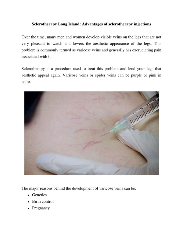
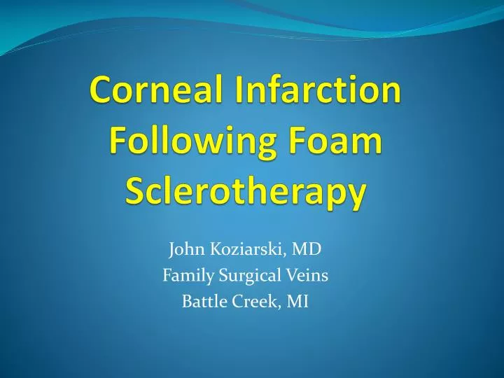
Corneal Infarction Following Foam Sclerotherapy. John Koziarski, MD Family Surgical Veins Battle Creek, MI. Financial Disclosures. I will be discussing the off-label use of medications. 2005 25 Year old Female. 6 yr history of pain, heaviness, aching since pregnancy 6 years prior

E N D
Corneal InfarctionFollowing Foam Sclerotherapy John Koziarski, MD Family Surgical Veins Battle Creek, MI
FinancialDisclosures I will be discussing the off-label use of medications
2005 25 Year old Female • 6 yr history of pain, heaviness, aching since pregnancy 6 years prior • No significant PMH • No hx of migraines
Ultrasound Exam • Bilat GSV reflux • Bilat SSV refluv • Incompetent Cockett perforator right leg • Bilat deep system insufficiency
Treatment Surgical Sclerotherapy • Thermal ablation bilat GSV and bilat SSV • Ligation incompetent Cockett perforator • Multiple sessions with 0.66% Polidocanol liquid
2009 Over the previous 1 yr Ultrasound • Increasing pain and aching and swelling left leg despite compression stockings • 5-6 mm reticular veins over thighs, 2-3 mm varicosities legs • No evidence of bilat GSV of bilat SSS • No neovascularity or refluxing acc veins • 3 incompetent perforators left thigh/leg • Bilat deep system reflux
Treatment Chemoablation Perf Sclerotherapy Superficial Varicosites • 3 perforators left thigh/leg treated • 0.5 ml of 2% STS/ CO2 foam (1:4) • Perivenous injection of NS to compress vein • Perforators closed • No complications
Jan 2010 Treatment of superficial varicosities • Left thigh • 2 ml 0.2% liquid STS • 4 ml 0.4% STS/CO2 foam (1:4) • Stocking applied • Pt went to work (in another physician’s office)
30 min post injection 60 min post injection • Called office c/o blurred vision left eye • Reassured that visual disturbances can happen and should resolve. • Said she would lie down for a while at work. • Called office again • Still blurred vision left eye • Now pain in right eye (10/10) and blurred vision rt eye • Nausea • BP 90/60 HR 70 SaO2 97%
To ER Chief Complaint Diagnostic Work Up • Blurred vision left eye resolved • Still had pain IN her rt eye and decreased vision • Headache • Chest pain/pressure • Nausea • No focal neurologic deficit • EKG • Troponin • CXR • MRI Brain • MRA/MRV brain • All Normal
ER Disposition Treated Discharged • Dilaudid • Zofran • Imitrex • No real improvement in pain, but nausea improved • Vicodin, Zofran • F/U Family Dr in am
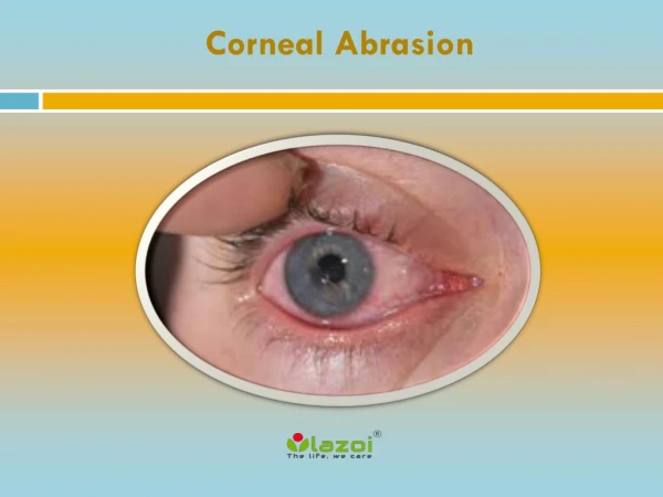
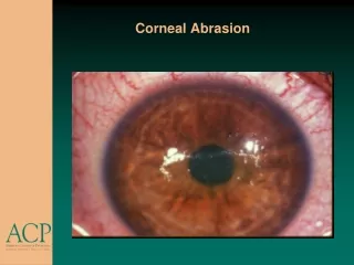
24 hours post injection Family Physician Ophthamologist • Main complaint right eye pain and vision loss • Some chest pain/pressure • Did not “seem” like a migraine • Later- CT scan chest- No PE • Vision right- 20/100 left- 20/20 • Retina normal • IOP normal • Rt cornea “ground glass” appearance • Ischemic?
Treatment • Eye patch • Drops • 6 Weeks • Cornea Healed • Visual acuity normal • Some light sensitivity
Further Workup • Transthoracic Echo • Small PFO • TEE with bubble study • No PFO • TCD with bubble study • No PFO
Further Workup Neurologist • Hypercoagulable w/u • Negative • Repeat MRI (Jun 2010) • Normal • Repeat MRI (Dec 2010) • Normal
Frequency of Visual Disturbances (VD) after Foam Sclerotherapy of 1.4% • Could be “positive” or “negative” or both • 50% headache • Other Sx included nausea, photophobia, chest pressure, and parathesias. • 18/20 pts with sx had Diffusion-Weighted MRI • All were normal. 5 had Non specific White Matter lesions
Hypothesizes that endothelin-1, released from the treated vessel endothelium, may be the mediator • VD can occur with liquid or foam, though more frequent with foam (Guex et al Dermatol Surg 2005) • Endothelin-1 has been associated with retinal vasospasm, migraine with aura, and bronchconstriction
Cornea • No blood supply • Receives O2 and nutrients from tears and aqueous humor • Endothelin-1 has been found in the Epithelium and tears (Lu et al.Exp Biol Med 2001) • Effects on Cornea are not well understood. May effect cell growth and apoptosis, and may promote corneal healing. (Salvatore, et al. J of Ophth 2010)
Summary • 30 yr old female, no history of migraine, underwent foam sclerotherapy of reticular veins with 4 ml of 0.4% STS/CO2 foam and 2 ml 0.2% STS liquid • Experienced what seemed to be “typical” visual disturbances, but with the addition of corneal injury • Etiology is unclear