Download
1 / 31
380 likes | 1.05k Views
Myocardial infarction. New concepts New definitions. Coronary Disease presentations. Angina Myocardial infarction Sudden cardiac death. Coronary Disease presentations. Angina stable unstable Myocardial infarction Sudden cardiac death. Coronary Disease presentations.

E N D
Myocardial infarction New concepts New definitions
Coronary Disease presentations • Angina • Myocardial infarction • Sudden cardiac death
Coronary Disease presentations • Angina stable unstable • Myocardial infarction • Sudden cardiac death
Coronary Disease presentations • Angina stable unstable • Myocardial infarction ‘full-thickness’, ‘transmural’, Q-wave ‘partial-thickness’, subendocardial’ • Sudden cardiac death
Coronary Disease presentations • Angina stable unstable • Myocardial infarction STEMI NSTEMI • Sudden cardiac death
Acute Coronary Syndrome No ST Elevation ST Elevation Unstable Angina NQMI QMI
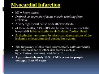
Traditional definition of MI • Symptoms of myocardial ischemia • Elevation of cardiac ‘enzymes’ in blood • Typical ECG patterns
New definitions of MI • Consensus document published in April 2000 by the American College of Cardiology and the European Society of Cardiology • Criteria for acute, evolving or recent MI • Criteria for established MI
Acute, evolving or recent MI • Typical rise and fall of biochemical markers of myocardial necrosis, CK-MB or Troponin associated with at least one of the following ischaemic symptomsnew pathological Q waves ECG changes indicative of ischaemiacoronary artery interventionor • Pathological findings of an acute MI
Established MI • Development of new pathological Q waves in serial ECGs or • Pathologic findings of a healed or healing MI
Cardiac biomarkers • CK-MB • Troponin T (or I) • Total CK, LDH and ASAT all invalid
CK-MB Improved sensitivity over earlier enzyme estimations All enzyme measurements have a background level of ‘noise’ - ie. normal range
CK-MB Improved sensitivity over earlier enzyme estimations All enzyme measurement have a background level of ‘noise’ - ie. normal range 1st detected 2 - 3hrs post MI, and elevation persists for 1 - 2 days
Troponin • = a protein (not an enzyme) • Troponin T and I (cardiac troponins) are not detectable in the blood of healthy subjects • Reliable lab test for T ; not for I • 1st detectable 3 – 4 hrs post MI, and persists for 7 – 14 days • Troponin T elevated (ie detectable) in conditions other than acute infarction
Coronary intervention Coronary artery spasm Coronary artery embolus Coronary artery inflammation Coronary artery dissection Direct coronary artery trauma Sympathomimetics Pulmonary embolus End-stage renal failure Rhythm disturbances Acute heart failure Extreme endurance exercise Secondary ischaemic cardiac injury
Non-ischaemic cardiac injury • Myocarditis – multiple causes • Cardiac trauma direct cardiac surgery • Metabolic / toxic renal failure
Clinical features • Spontaneous ischaemic episode, usually lasting > 20 minutes • Coronary artery intervention angiography, angioplasty, stenting
ECG features of myocardial ischaemia that may MI • New ST elevation in at least two contiguous leads, measuring >= 0.2mV in leads V1 – V3, or >= 0.1mV in all other leads = STEMI • Absence of ST elevation, but with either ST depression or T wave abnormalities = NSTEMI
ECG features of established MI • In absence of confounders (LBBB, LVH, and WPW syndrome) any Q wave in V1 – V3, or Q waves of >= 1mm for >= 30msec in two other contiguous leads
Normal ECG Normal ST segment Normal Q wave ST elevation ST depression Pathological Q wave
pathology • 6 hours elapse before myocardial necrosis becomes evident on histopathology • Three phases – acute / healing / healed • Size – microscopic / small / medium / large
imaging • Echocardiography • Radionuclide angiography • Single-photo emission computed tomography = SPECT
Key points in the new definitions of myocardial infarction • Any myocardial necrosis constitutes an infarction • Infarctions other than ‘spontaneous’ are included
Specific clinical situations not yet defined • MI post-CABG, based on post-operative CK-MB or troponin levels • Threatened and aborted infarction • Silent infarction • Sudden ischaemic cardiac death
Myocardial Infarction Redefined - A Consensus Document of the Joint European Society of Cardiology / American College of Cardiology Committee for the Redefinition of Myocardial Infarction Journal of the American College of Cardiology 36:959-969 2000