Download
1 / 27
380 likes | 1.11k Views
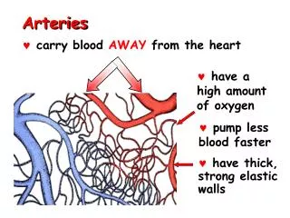
Basic Definitions. Arteries carry blood away from the heart Veins carry blood back to the heart Arterioles = small arteries Venules = small veins

E N D
Basic Definitions • Arteries carry blood away from the heart • Veins carry blood back to the heart • Arterioles = small arteries • Venules = small veins • Capillaries: smallest vessels where nutrient, waste and gas exchange take place. So narrow that blood cells proceed through single file. Walls are a single layer of epithelium.
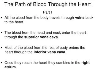
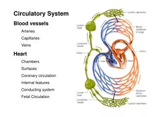
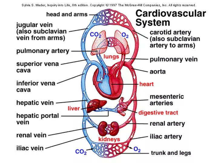
Summary Concepts • The human heart has four chambers left and right atria, left and right ventricles • The human heart has four valves two atrioventricular valves (tricuspid and mitral) two semilunar valves (aortic semilunar and pulmonary semilunar) • Pulmonary circulation: starts as blood leaves the right ventricle and enters the pulmonary trunk -> lungs -> pulmonary veins -> left atrium • Systemic circulation: starts as blood leaves the left ventricles -> aorta -> head, arms, legs, body -> vena cava -> right atriumCoronary circulation: provides blood to the heart muscle (myocardium). Aorta -> coronary arteries -> heart muscle -> cardiac veins -> vena cava.
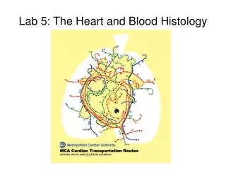
Subclavian artery Left carotid artery Superior vena cava Brachiocephalic artery Aortic arch Pulmonary arteries Pulmonary veins Pulmonary trunk left atrium Right Atrium Coronary arteries Cardiac veins Left Ventricle Right ventricle Inferior vena cava
Atria: receive blood from principle veins and assist in filling the ventricles Ventricles: pump blood under high pressure into pulmonary trunk and the aorta. Left Atrium Aortic Semilunar Right Atrium Pulmonary Semilunar Bicuspid (mitral) Tricuspid Left Ventricle Right Ventricle
Tissues of the Heart • Pericardium: serous membrane sack surrounding the heart • Epicardium: serous membrane on outer surface of the heart • Myocardium: muscle of the heart • Endocardium: membrane lining the interior of the heart • Chordae tendonae: “heart strings” that guide the atrioventricular valves. • Papillary muscle: small muscles that anchor the chordae tendonae to the ventricular walls.
Heart Valves are passive and move According to pressure gradients (like a loose screen door in a breeze) Atrioventricular Semilunar Aorta Superior Vena Cava 8 7 Pulmonary trunk 4 Pulmonary Vein 3 5 1 6 2 Inferior Vena Cava video
The Cardiac Cycle Diastole: period of relaxation And ventricular filling Atrial Systole: atria contract force additional blood into the ventricles. Ventricular Systole: ventricles contract and force blood into pulmonary trunk and aorta.
Purkinje Fibers Conduction System Video
Electrical Recovery of Myocardium R T P Q S Electrocardiogram
Cardiac Output Stroke Volume (mL/beat) X Heart Rate (beats/min) = Cardiac Output (mL/min) 75 mL/beat X 72 beats/min. = 5400 mL/min. • Questions • Do athletes have greater or lesser demands for oxygen than non-athletes? • Do athletes have greater or lesser resting heart rates than non-athletes? • How do you reconcile this observation? Affects of Athletic Training - Stroke volume increases, heart rate decreases 95 mL/beat X 60 beats/min. = 5700 mL/min. Even though the athlete has a lower resting heart rate, s/he has a greater Cardiac Output than the untrained person.
Cardiac Center in the Medulla Controls two nerves that lead to the heart1. accelerator nerve (sympathetic) *acts on the sinoatrial node and ventricles *increases heart rate using norepinephrine2. vagus nerve (parasympathetic) *acts on the sinoatrial node *decreases heart rate using acetylcholine
Regulation of Heart Rate • Sensory Inputs • Proprioceptors(Am I moving more?) • Baroreceptors(Is my blood pressure low?) • Chemoreceptors(Is my CO2 high or O2 low?) Cardiac Center(medulla) Vagus nerve Sensory nerves Proprioceptors • Motor Output • Accelerator nerve(sympathetic)(releases norpepinephrine)increase rate and force • Vagus nerve(parasympathetic)decrease rate Acceleratornerve Baroreceptors and Chemoreceptors in the carotid and aortic arteries
Arteries = elastic elements Veins = capacitance elements compliant valves skeletal muscles
Arterioles and pre-capillary Sphincters are muscular constrict add resistance dilate reduce resistance Large arteries are elastic absorb kinetic energy store potential energy Each organ has its own set of Arterioles, and capillary beds Which may be dilated or constricted
The microcirculation 1. Capillary exchange by diffusion Vitamins nutrients CO2 wastes O2
Capillary exchange by bulk flow • Starling hypothesis • - capillaries have low permeability to protein (osmotic effect) • - hydrostatic pressure decreases as blood passes through Net outward pressure = 11 mmHg Net inward pressure = -9 mmHg
Edema • Alcoholism • Elephantiasis