Download
1 / 58
640 likes | 1.23k Views
Heart and Blood Vessels. 0. 8. Blood Vessels Transport Blood. Arteries Carry blood away from the heart Transport blood under high pressure Thick-walled Capillaries Exchange solutes and water with cells of the body Microscopic Veins Return blood to the heart Thin-walled. Figure 8.1.

E N D
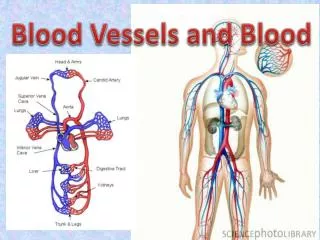
Blood Vessels Transport Blood Arteries Carry blood away from the heart Transport blood under high pressure Thick-walled Capillaries Exchange solutes and water with cells of the body Microscopic Veins Return blood to the heart Thin-walled
Figure 8.1 Direction of blood flow Outer layer: Connective tissue Middle layer: Smooth muscle with elastic fibers Inner layer: Endothelium Vein Artery Connective tissue Smooth muscle Endothelium Arteriole Venule Capillary Epithelial cells of capillary endothelium Tissue cells
Arteries Transport Blood Away From Heart Structure Thick-walled, three layers Innermost layer: endothelium of squamous epithelial cells Middle layer: smooth muscle Outer layer: connective tissue Function Arteries carry blood away from heart Carry blood under pressure
Arteries Transport Blood Away From the Heart Aneurism—defect in arterial wall Ballooning of the arterial wall Some bulge inward, obstructing flow Others bulge outward Often develop slowly over time Often symptomless, until they rupture Rupture of aortic aneurism—rapidly fatal May be detected by careful screening and surgically repaired
Arterioles and Precapillary Sphincters Regulate Blood Flow Blood flow Heart Arteries Arterioles Capillaries Arterioles: smallest arteries Precapillary sphincters: control blood flow from arterioles into capillaries Vasodilation: Relaxation of vascular smooth muscle Increases blood flow to capillaries Vasoconstriction: Contraction of vascular smooth muscle Decreases blood flow to capillaries
Figure 8.2 Constricted precapillary sphincters Relaxed precapillary sphincters Arteriole Capillaries Small vein (venule)
Capillaries: Where Blood Exchanges Substances with Tissues Structure Smallest blood vessels, microscopic Thin-walled: one cell-layer thick Porous Capillary beds: extensive networks of capillaries Function: selective exchange of substances with the interstitial fluid
Figure 8.3 Capillary cell Pores through cells Slit between cells A medium-magnification view showing a rich network of capillaries surrounding and interconnecting small arteries and veins. Nucleus RBC A higher magnification showing a single branching capillary. Notice the red blood cells traveling single file in the capillary. The structure of a capillary.
Figure 8.4 Precapillary sphincter Capillary RBCs, most proteins Fluid (water) O2, nutrients, raw materials CO2, wastes Venule Arteriole Tissue cell
Lymphatic System Helps Maintain Blood Volume Function Maintains blood volume Returns excess interstitial fluid to circulatory system Also functions in immune defenses Structure Blind-ended capillaries Lymphatic vessels (similar to venous system) Lymph—derived from interstitial fluid
Veins Return Blood to the Heart Structure Three layers, thin-walled Larger lumen than arteries High distensibility Functions Carry blood toward the heart Blood flow Capillaries Venules Veins Heart Serve as blood volume reservoir
Veins Return Blood to the Heart Three Mechanisms assisting in blood return Contraction of skeletal muscles One-way valves permit only one-way blood flow Pressure changes associated with breathing push blood toward the heart
Figure 8.5 Valve (open) One-way valves (closed) Calf muscles relaxed Calf muscles contracted
Figure 8.7 Aorta Superior vena cava Left pulmonary artery Right pulmonary artery Pulmonary trunk Left pulmonary veins Left atrium Pulmonary semilunar valve Aortic semilunar valve Left atrioventricular (AV) valve Right atrium Left ventricle Right atrioventricular (AV) valve Chordae tendineae Papillary muscles Septum Right ventricle Epicardium Inferior vena cava Myocardium Endocardium
The Heart Is Mostly Muscle Surrounded by fibrous sac—pericardium Protects and anchors the heart Layers of the heart Epicardium: thin layer of epithelial and connective tissue Myocardium: thick layer of cardiac muscle Electrical signals flow directly from cell to cell This is what contracts when the heart beats Endocardium: thin layer of endothelial tissue Continuous with lining of blood vessels
The Heart Has Four Chambers and Four Valves Four chambers Two atria: upper chambers Two ventricles: lower chambers Septum, muscular partition separates right and left sides of the heart Four valves—prevent backflow Two atrioventricular (AV) valves Tricuspid valve (right side) Bicuspid (mitral) valve (left side) Two semilunar valves Pulmonary valve Aortic valve
Animation: The Cardiovascular System Right-click and select Play
The Pulmonary Circuit Provides for Gas Exchange Deoxygenated blood from the body travels through the vena cava to the right atrium Through the right AV valve into the right ventricle Through the pulmonary semilunar valve into the pulmonary trunk and the lungs Blood is oxygenated and CO2 is given up within pulmonary capillaries Oxygenated blood travels through the pulmonary veins into the left atrium Through the left AV valve into the left ventricle
The Systemic Circuit Serves the Rest of the Body Oxygenated blood travels from the left ventricle through the aortic semilunar valve into the aorta Through branching arteries and arterioles to tissues Through the arterioles to capillaries Within capillaries, nutrients and oxygen are delivered and wastes are picked up From capillaries into venules and veins To the vena cava and into the right atrium
Figure 8.8 Systemic Circuit Head and upper limbs Pulmonary Circuit Lung capillaries Lung capillaries Aorta Heart Torso and Lower limbs
Figure 8.9 Jugular vein Subclavian vein Carotid artery Subclavian artery Superior vena cava Inferior vena cava Aorta Renal artery Renal vein Common iliac vein Femoral vein Femoral artery Common iliac artery Great saphenous vein
The Systematic Circuit Serves the Rest of the Body Coronary arteries Arteries that supply the heart muscle itself Supply the myocardium Small diameter—may become partially or completely blocked by atherosclerosis
Figure 8.10 Aorta Superior vena cava Pulmonary trunk Cardiac vein Left coronary artery Right coronary artery Cardiac veins Inferior vena cava
The Cardiac Cycle: The Heart Contracts and Relaxes Atrial systole Both atria contract AV valves open, semilunar valves are closed Ventricles fill Ventricular systole Both ventricles contract AV valves close, semilunar valves open Diastole Both atria and ventricles relax Semilunar valves close
Figure 8.11 Left atrium Right atrium Aortic semilunar valve Pulmonary semilunar valve Left AV valve Right AV valve Left ventricle Atrial systole. Both atria contract, forcing blood into the ventricles. The AV valves are open, and the semilunar valves are closed. Right ventricle 0.1 second Systole Diastole 0.4 second Aorta Pulmonary trunk 0.3 second Diastole. The ventricles relax and begin to fill passively with blood through the open AV valves. The semilunar valves are closed, and the atria remain relaxed. Ventricular systole. Both ventricles contract, causing the AV valves to close and the semilunar valves to open. Blood is ejected into the pulmonary trunk and aorta. The atria relax.
Heart Sounds Reflect Closing Heart Valves Lub-dub heart sound Lub: closing of both AV valves during ventricular systole Dub: closing of both semilunar valves during ventricular diastole Heart murmurs Caused when blood flow is disturbed May be a sign of a defective valve
Cardiac Conduction System Coordinates Contraction Sinoatrial (SA) node—small mass of cardiac cells in upper right atrium Cardiac pacemaker Initiates the heartbeat spontaneously Pace can be modified by nervous system Atrioventricular (AV) node Located between atria and ventricles Relays impulse Atrioventricular (AV) bundle and Purkinje fibers Located in septum and ventricles Carry impulse to ventricles
Figure 8.13 Sinoatrial (SA) node Atrioventricular (AV) node AV bundle Bundle branches Purkinje fibers
Electrocardiogram (EKG/ECG) Records the Heart’s Electrical Activity Tracks the electrical activity of the heart A healthy heart produces a characteristic pattern Three formations P wave: impulse across atria QRS complex: spread of impulse down septum, around ventricles in Purkinje fibers T wave: end of electrical activity in ventricles EKGs can detect Arrhythmias Ventricular fibrillation
Figure 8.14 An ECG being recorded. R T P Q S A normal ECG recording. Ventricular fibrillation.
Blood Exerts Pressure Against Vessel Walls The force that the blood exerts on the wall of the blood vessels Systolic pressure: highest pressure, as blood is ejected during ventricular systole Diastolic pressure: lowest pressure, during ventricular diastole Measurement Sphygmomanometer: device used to measure blood pressure “Normal” readings Systolic pressure <120 mmHg Diastolic pressure <80 mmHg
Figure 8.15 Systolic pressure 120 Blood pressure (mm Hg) 80 Diastolic pressure 40 0 Veins Arteries Venules Arterioles Capillaries
Figure 8.16 140 Cuff pressure Blood pressure 1 120 100 Blood pressure (mm Hg) Column of mercury indicating pressure in mm Hg 80 2 60 4 6 10 2 8 0 Sphygmomanometer: Time (seconds) A schematic representation of the pulses of arterial blood pressure superimposed over the steadily declining cuff pressure. Systolic pressure is recorded at cuff pressure 1 when sounds are first heard. Diastolic pressure is recorded at cuff pressure 2 when sounds cease. Squeezable bulb Inflatable rubber cuff Air valve Artery Stethoscope A clinician inflates the cuff with air and then allows the pressure in the cuff to fall gradually while using a stethoscope to listen for the sounds of blood movement through the artery.
Hypertension: High Blood Pressure Can Be Dangerous Sustained elevation in blood pressure Systolic pressure 140 mmHg Diastolic pressure 90 mmHg Risk factor for cardiovascular disease Higher blood pressure causes greater strain on cardiovascular system Blood vessels react by becoming hardened and scarred Strain on heart from having to work harder Silent killer, no symptoms
Hypotension: When Blood Pressure Is Too Low Low blood pressure If low enough, may cause dizziness or fainting May follow abrupt changes in body position Standing up suddenly May result from excessive blood loss or fluid loss from burns
How the Cardiovascular System Is Regulated Importance of maintaining a constant arterial blood pressure Constant arterial pressure is achieved by Regulation of heart rate Force of contraction Regulation of diameter of arterioles Local blood flows are adjusted to meet local requirements
Baroreceptors Maintain Arterial Blood Pressure Baroreceptors: pressure receptors in aorta and carotid arteries Steps in mechanism Blood pressure rises, vessels stretched Signals sent to the cardiovascular center in the brain Heart signaled to lower heart rate and force of contraction Arterioles vasodilate, increasing blood flow to tissues Combined effect lowers blood pressure Mechanism reversed if blood pressure is too low
Nerves and Hormones Adjust Cardiac Output Amount of blood pumped into aorta in one minute Cardiac output heart rate stroke volume Heart rate: beats/minute (bpm) Resting adult heart rate approx. 75 bpm Stroke volume: Resting adult stroke volume 70 ml/beat Resting cardiac output 75 bpm 70 ml/beat 5.25 liters/min.
Nerves and Hormones Adjust Cardiac Output Medulla oblongata: cardiovascular center of brain Receives inputs from baroreceptors and other receptors Output goes through two sets of nerves 1. Sympathetic nerves—constrict blood vessels, raising blood pressure 2. Parasympathetic nerves—dilate blood vessels, lowering blood pressure Hormones: epinephrine and norepinephrin Secreted by adrenal glands when sympathetic system is activated Increase cardiac output
Local Requirements Dictate Local Blood Flows Precapillary sphincters allow fine-tuning of blood flow to local tissues as needed Metabolically active tissue—needs more O2, sphincters open, vasodilation If blood pressure drops precipitously, blood pressure control would cause vasoconstriction to many organs and shunt blood to brain and heart where blood supply and pressure must be maintained
Figure 8.17 Blood flow Cell Diffusion of vasodilating substance Arteriole Precapillary sphincter Capillary Vasodilating substance produced during metabolism At rest, very little of the vasodilating substance would be produced, and flow would be minimal. With increased metabolic activity, the presence of more of the substance in the interstitial space would cause the arteriole and precapillary sphincter to vasodilate, increasing flow.
Exercise: Increased Blood Flow and Cardiac Output Blood flow to active skeletal muscles increases Cardiac output (CO) is increased to maintain blood pressure Non-athletes: up to 20–25 liters/min Trained athletes: up to 35 liters/min
Cardiovascular Disorders: A Major Health Issue Angina Sensation of pain and tightness in chest Caused by narrowing of coronary arteries and diminished blood flow to coronary muscle May be accompanied by shortness of breath and sensation of choking or suffocating Usually temporary Angiography: allows visualization of coronary arteries, enables diagnosis of angina Treatment: Medication Balloon angioplasty Coronary artery bypass graft
Cardiac Disorders: A Major Health Issue Heart attack (myocardial infarction) Sudden death of an area of myocardium Symptoms: Intense chest pain, tightness or pressure on chest, radiating left arm pain, jaw and back pain, nausea Requires immediate medical attention Diagnosis: ECG and presence of certain enzymes in the blood Treatment and/or prevention Control of arrhythmias Clot-dissolving medications Coronary artery bypass graft (CABG)—vein from leg is grafted to bypass obstructed coronary artery
Figure 8.19 Aorta Vein grafts Plaque blocking blood flow