Download
1 / 37
410 likes | 608 Views
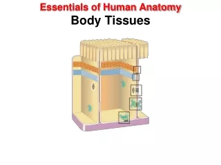
Body Tissues. Ch.3 (Part II). Body Tissues. Cells are specialized for particular functions Tissues Groups of cells with similar structure and function Four primary types Epithelium Connective tissue Nervous tissue Muscle. Body Tissues.

E N D
Body Tissues Ch.3 (Part II)
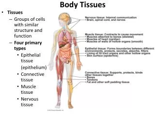
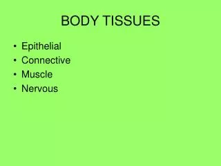
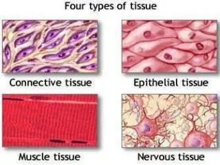
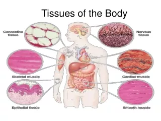
Body Tissues • Cells are specialized for particular functions • Tissues • Groups of cells with similar structure and function • Four primary types • Epithelium • Connective tissue • Nervous tissue • Muscle
Body Tissues • If we had to assign a single term to each primary tissue type that would best describe its overall role, the terms would most likely be: • Covering (epithelium) • Support (connective) • Movement (muscle) • Control (nervous) • However, these terms only reflect only a tiny fraction of the functions that each of these tissues perform.
1. Epithelial Tissues • Found in different areas • Body coverings • Body linings • Glandular tissue (forms glands in body) • Functions • Protection • Absorption • Filtration • Secretion
Special Characteristics of Epithelium: • Cells fit closely together (except for glandular) to form connective sheets • Tissue layer always has one free surface. This so called “apical surface” is exposed to the body’s exterior or to the cavity of an internal organ. • The lower surface of epithelium is bound by a basement membrane (a structure less material secreted by cells) • Avascular (have no blood supply) so they depend on diffusion from the capillaries in the underlying connective tissue for food and oxygen • Regenerate easily if well nourished
Classification of Epithelium 1. Number of cell layers • Simple – one layer • Stratified – more than one layer Figure 3.17a
Classification of Epithelium 2. Shape of cells • Squamous – flattened (like fish scales) • Cuboidal – cube-shaped (like dice) • Columnar – column-like (like columns…duh) Figure 3.17b
Simple or Stratified… Simple Stratified Consist of two or more cell layers More durable than the simple epithelia Function primarily to protect • One layer of cells • Most concerned with absorption, secretion, and filtration • Because they are usually very thin, protection is not one of their specialties
Simple Epithelium • 1. Simple squamous • Single layer of flat cells • Usually forms membranes where filtration or exchange of substances by rapid diffusion occurs. • Lines body cavities • Lines lungs and capillaries Figure 3.18a
Simple Epithelium • 2. Simple cuboidal • Single layer of cube-like cells • Common in glands and their ducts • Forms walls of kidney tubules • Covers the ovaries Figure 3.18b
Simple Epithelium • 3. Simple columnar • Single layer of tall cells • Often includes goblet cells, which produce mucus • Lines the entire digestive tract from the stomach to the anus Figure 3.18c
Simple Epithelium • 4. Pseudostratified • Single layer, but some cells are shorter than others • Often looks like a double cell layer (false…hence the name pseudo) • Sometimes ciliated, such as in the respiratory tract • May function in absorption or secretion Figure 3.18d
Stratified Epithelium • 1. Stratified squamous • Cells at the free edge are flattened but closer to the basement membrane, are cuboidal or columnar • Found as a protective covering where friction/abuse is common • Locations • Skin • Mouth • Esophagus Figure 3.18e
Stratified Epithelium • 2. Stratified cuboidal • Two layers of cuboidal cells • 3. Stratified columnar • Surface cells are columnar, cells underneath vary in size and shape • Stratified cuboidal and columnar • Rare in human body • Found mainly in ducts of large glands
Stratified Epithelium • 4. Transitional epithelium • Shape of cells depends upon the amount of stretching (near basement: typically columnar or cuboidal, at surface: varies) • Lines organs of the urinary system like the bladder, ureters, and part of the urethra where considerable amounts of stretching occur. Figure 3.18f
Glandular Epithelium • Gland – one or more cells that secretes a particular product (secretion) • Two major gland types develop from epithelial sheets: • 1. Endocrine gland • Ductless (hormones diffuse directly into the blood vessels that weave through the glands) • Secretions are all hormones • Examples of endocrine glands: thyroid, adrenals, and pituitary • 2. Exocrine gland • Empty through ducts to the epithelial surface • Include sweat and oil glands, and liver and pancreas • Are both internal and external
Connective Tissue • Found everywhere in the body & connects body parts • Includes the most abundant and widely distributed tissues • Functions • Binds body tissues together • Supports the body • Provides protection
Connective Tissue Characteristics: • Variations in blood supply • Some connective tissue types are well vascularized (good blood supply) • Some have poor blood supply or are avascular like tendons and cartilages (these things heal slowly) • Extracellular matrix • Connective tissues are made up of many different types of cells plus varying amounts of non-living material that surrounds living cells (called the extracellular matrix)
Extracellular MatrixMakes connective tissue different from any other tissue. • Two main elements • Ground substance – mostly water along with adhesion proteins and polysaccharide molecules • Fibers • Produced by the cells • Three types • Collagen fibers • Elastic fibers • Reticular fibers
Extracellular Matrix Continued: • Allows connective tissue to • form a soft packaging tissue around other organs, • to bear weight, • and to withstand stretching and other abuses, such as abrasion that no other tissue could endure.
Connective Tissue Types: • Bone (osseous tissue) • Composed of: • Bone cells in lacunae (cavities) • Surrounded by a hard matrix of calcium salts • Large numbers of collagen fibers • Because of its rocklike hardness, bone is used to protect and support the body Figure 3.19a
Connective Tissue Types: • Hyaline cartilage • Most common cartilage (cartilage is found only in a few places in the body…is less hard and more flexible than bone.) • Composed of: • Abundant collagen fibers • Rubbery matrix • Blue-white appearance • Entire fetal skeleton is hyaline cartilage (turns to bone by birth)! • Also forms the supporting structure of the larynx (voicebox), attaches ribs to the breastbone, and covers the ends of bones where they form joints. Figure 3.19b
Connective Tissue Types: • Elastic cartilage • Provides elasticity • Example: supports the external ear
Connective Tissue Types: • Fibrocartilage • Highly compressible • Example: forms cushion-like discs between vertebrae Figure 3.19c
ConnectiveTissue Types: • Dense connective tissue • Main matrix element is collagen fibers • Cells are fibroblasts (fiber forming cells) • Forms strong ropelike structures (like the examples listed below) • Examples • Tendon – attach muscle to bone • Ligaments – attach bone to bone Figure 3.19d
Connective Tissue Types: • Areolar connective tissue • Most widely distributed connective tissue • Soft, pliable “cobwebby” tissue that cushions and protects the body organs • Helps hold internal organs together and in their proper positions • Contains all fiber types • Can soak up excess fluid Figure 3.19e
ConnectiveTissue Types: • Adipose tissue: Commonly called fat! • Matrix is an areolar tissue in which fat cells predominate • Many cells contain large lipid deposits • Functions • Insulates the body • Protects some organs • Serves as a site of fuel storage Figure 3.19f
Connective Tissue Types: • Reticular connective tissue • Delicate network of interwoven fibers • Forms stroma (internal supporting network) of lymphoid organs • Lymph nodes • Spleen • Bone marrow Figure 3.19g
Connective Tissue Types: • Blood (vascular tissue) • Blood cells surrounded by fluid matrix (called plasma) • Fibers are visible during clotting • Functions as the transport vehicle for the cardiovascular system, carrying nutrients, wastes, respiratory gases, and many other substances throughout the body. Figure 3.19h
Muscle Tissue • Function is to produce movement • Three types • Skeletal muscle • Cardiac muscle • Smooth muscle
Muscle Tissue Types: • 1. Skeletal muscle • Can be controlled voluntarily • Form the muscular system • Cells attach to connective tissue • Cells are striated (striped) • Cells have more than one nucleus Figure 3.20a
Muscle Tissue Types • 2. Cardiac muscle • Found only in the heart • Function is to pump blood (involuntary) • Cells attached to other cardiac muscle cells at intercalated disks (have gap junctions …remember this mean info can pass freely from cell to cell….) • Cells are striated • One nucleus per cell • Under involuntary control Figure 3.20b
Muscle Tissue Types • 3. Smooth muscle • AKA: visceral muscle • Involuntary muscle • Surrounds hollow organs • Spindle-shaped (pointed at each end) • Attached to other smooth muscle cells • No visible striations (hence the name smooth) • One nucleus per cell Figure 3.20c
Nervous Tissue • Neurons and nerve support cells • Function is to send impulses to other areas of the body • Irritability • Conductivity Figure 3.21
Tissue Repair (Wound Healing) • Tissue repair occurs in two major ways… • 1. Regeneration • Replacement of destroyed tissue by the same kind of cells • 2. Fibrosis • Repair by dense fibrous connective tissue (scar tissue is formed) • Determination of method • Type of tissue damaged • Severity of the injury
Tissue injury sets a series of events into motion… • Capillaries become very permeable • Introduce clotting proteins • Wall off injured area • Formation of granulation tissue • Regeneration of surface epithelium
Regeneration of Tissues • Tissues that regenerate easily • Epithelial tissue • Fibrous connective tissue and bone • Tissues that regenerate poorly • Skeletal muscle • Tissues that are replaced largely with scar tissue • Cardiac muscle • Nervous tissue within the brain and spinal cord Read about Homeostatic Imbalance on pg. 98 and make notes….