Download
1 / 46
480 likes | 659 Views
Body Tissues. Overview. Human body starts with one cell Division makes millions of cells Each specialized for particular functions Some are so specialized, they can create hazards Ex. Heart cells Groups of similar cells that are similar in structure and function: tissues. Types of Tissues.

E N D
Overview • Human body starts with one cell • Division makes millions of cells • Each specialized for particular functions • Some are so specialized, they can create hazards • Ex. Heart cells • Groups of similar cells that are similar in structure and function: tissues
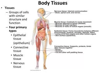
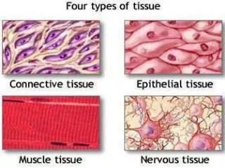
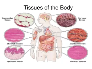
Types of Tissues • Epithelium (covering) • Connective (support) • Nervous (control) • Muscle (movement) • All interweave to form the fabric of the body • Tissues organized into organs • One organ can be composed of several tissue types
Epithelial Tissue • Also called epithelium • Lining, covering, and glandular • Forms glands • Covers all free body surfaces • Versatile cells • Outer skin layer, and lining of body cavities • Every substance entering or leaving must pass through these cells
Special Characteristics • Except for glandular, they fit closely together to form continuous sheets • Always have one free surface (apical surface): exposed to body’s exterior or the cavity of an internal organ • Lower surface rests on a basement membrane, structureless material secreted by cells • No blood supply of their own (avascular); depend on diffusion from capillaries • Regenerate themselves very easily
Classification of Epithelium • Two names • First name is for number of cells: simple (one) or stratified (more than one) • Second name describes shape: squamous (flattened), cuboidal (cube-shaped), and columnar (column-shaped) • Names are combined • *Stratified epithelia are named for the cells at the free surface of the membrane, not the those resting on the basement membrane
Simple Epithelia • Most concerned with absorption, secretion, and filtration • Simple Squamous epithelium • Single layer resting on basement membrane • Fit closely together • Usually forms membranes that use diffusion: air sacs of lungs, walls of capillaries • Also forms serous membranes or serosae: slick membranes lining ventral body cavity and cover organs in ventral cavity
Simple Epithelia contd. • Simple CuboidalEpi. • One layer resting on basement membrane • Common in glands and ducts: salivary glands and pancreas • Forms walls of the kidney tubules, and covers surface of the ovaries
Simple Epithelia contd. • Simple Columnar Epi. • One layer of tall cells • Fit close together • Goblet cells: produce a lubricating mucus, often seen in this type of epithelium • Lines entire length of digestive tract from stomach to anus • *Epithelial membranes that line body cavities open to the exterior are called mucous membranes or mucosae
Simple Epithelia contd. • Pseudostratified Columnar Epi. • Rest on basement membrane • Cells are different heights, and nuclei appear at different heights above basement • Give false (pseudo) impression of stratified • Mainly absorption and secretion • Pseudostratified ciliated columnar epithelium • Ciliated, lines most of respiratory tract • Goblet cells produce mucus to trap dust and debris
Stratified Epithelia • Two or more cell layers • More durable than simple • Primarily for protection • Stratified Squamous Epithelium • Most common stratified • Usually several layers • Free edge are squamous, closer to basement are cuboidal or columnar • Found in “high friction” areas: esophagus, mouth, other parts of skin
Stratified Epithelia • Stratified Cuboidal and Columnar Epithelia • Usually just two cell layers with (at least) the surface cells being cuboidal • Surface cells of stratified columnar are columnar, but its basal cells vary in size and shape • Both are fairly rare • Found mainly in the ducts or large glands
Stratified Epithelia • Transitional Epithelium • Highly modified • Stratified squamous epithelium • Forms lining of only a few organs: bladder, the ureters, and part of the urethra • All part of the urinary system and undergo considerable stretching • Basal layer are cuboidal or columnar; those at the free surface vary in appearance • Stretching changes shape, cells can flatten and become squamous-like
Glandular Epithelia • Gland: consists of one more cells that make and secrete a particular product • Secretion: typically contains protein molecules in an aqueous fluid • Noun and verb: glandular cells obtain needed materials from the blood and use them to make their secretion, which they then discharge • Two types: • Endocrine glands: no connection to surface (ductless), secrete hormones through diffusion • Exocrine glands: retain ducts, secretions empty to surface: sweat and oil glands, liver and pancreas, internal and external
Connective Tissue • Connects body parts • Found everywhere in body • Most abundant and widely distributed • Protect, support, bind other tissues together • Most are well vascularized (good blood supply) • Exceptions: tendons and ligaments have poor and cartilage has none (tend to heal slower than the others) • Extracellular matrix: nonliving substance found outside of these cells
Extracellular Matrix • Only found in connective tissue • Produced by the connective tissue cells then secreted to their exterior • Two main elements: • Structureless ground substance: water + adhesion proteins and large charged polysaccharide molecules; like the “glue” between these cells • Fibers: collagen (white) very strong; elastic (yellow) stretching; reticular (fine collagen fibers) make up internal “skeleton” of soft organs such as spleen
Extracellular Matrix • Because of matrix, connective tissue is able to form a soft packing tissue around other organs, to bear weight, and to withstand stretching and other abuses • Varies depending on tissue type: • Fat tissue is mostly cells and the matrix is soft • Bone and cartilage have very few cells and large amounts of hard matrix which makes them strong
Types of Connective Tissue • From most rigid to softest: • Bone • Cartilage • Dense Connective tissue • Loose Connective tissue • Blood
Bone • Called Osseous tissue • Composed of bone cells sitting in cavities called lacunae • Surrounded by layers of a very hard matrix that contains calcium salts and collagen fibers • Exceptional ability to protect and support
Cartilage • Less hard and more flexible than bone • Found only in a few places • Most widespread is hyaline cartilage: lots of collagen fibers, glassy, blue-white appearance • Forms larynx, attaches ribs to breastbone, covers the ends of bones; skeleton of fetus • Fibrocartilage: highly compressible, forms disks between the vertebrae of spinal column • Elastic Cartilage:found where elasticity is desired; external ear
Dense Connective Tissue • Also called dense fibrous tissue • Collagen fibers as its main matrix element • Between collagen fibers are rows of fibroblasts (fiber-forming cells) • Forms strong, rope-like structures such as tendons (attach skeletal muscles to bones) and ligaments (connect bones to bones at joints) • Also makes up lower layers of skin (dermis), arranged in sheets
Loose Connective Tissue • Softer and have more cells and fewer fibers • 3 types: • Areolar: most widely distributed of connective; soft, pliable, “cobwebby”; cushions and protects; universal packing tissue; holds internal organs together and in place; lamina propriaunderlies all mucous membranes; all types of fibers forming loose network; provides reservoir of water and salts for surrounding tissues; inflammation in the body causes this tissue to soak up excess fluid like a sponge which causes it become puffy: edema; phagocytes wander through looking for debris to destroy
Adipose Tissue • Commonly called fat • Areolar with fat cells dominate • Fat cells = signet ring cells: cells are full of oil – pushes nucleus to outer rim of cell • Forms subcutaneous tissue beneath skin • Insulates and protects from heat and cold • Protects some organs: kidneys, eyeballs in sockets • Fat depots in body: hips and breasts where fat is stored and available for energy if needed
Reticular Tissue • Delicate network of interwoven reticular fibers associated with reticular cells • Forms the stroma: internal framework, which can support many free blood cells (mostly lymphocytes) in lymphoid organs such as lymph nodes, the spleen, and bone marrow
Blood • Vascular tissue • Consists of blood cells, surrounded by nonliving, fluid matrix called blood plasma • “fibers” in blood are soluble proteins that only become visible during clotting • Transport vehicle for the cardiovascular system • Carries nutrients, wastes, respiratory gases, and many other substances
Muscle Tissue • Highly specialized to contract or shorten • Produce movement • Three types: • Skeletal muscle • Cardiac muscle • Smooth muscle
Skeletal Muscle • Packaged by connective tissue sheets into organs called skeletal muscles, which are attached to the skeleton • Controlled voluntarily (consciously) • Form the flesh of the body: the muscular system • When they contract they pull on bones or skin • Cells are long, cylindrical, multinucleate, and striations (stripes) • Often called muscle fibers
Cardiac Muscle • Found only in heart • Has striations • Uninucleate • Short • Branching cells that fit tightly together at junctions called intercalated discs • Allow for rapid conduction of electrical impulses • Controlled involuntarily
Smooth Muscle • Also called visceral muscle • No striations visible • Single nucleus, and spindle shaped (pointed at ends) • Walls of hollow organs: stomach, bladder, uterus, and blood vessels • Contracts slower than other types • Peristalsis: wavelike motion that keeps food moving through small intestine
Nervous Tissue • Cells called neurons • Receive and conduct electrochemical impulses from one body part to another • Irritability and conductivity are major functions • Unique structure • Cytoplasm drawn out into long processes (extensions) as much as 3 ft or more in the leg • Supporting cells: insulate, support, and protect the delicate neurons • Make up nervous system: brain, spinal cord, and nerves