Download
1 / 80
820 likes | 848 Views
Body Tissues. Levels of Structural Organization. Chemical – atoms combined to form molecules Cellular – cells are made of molecules Tissue – consists of similar types of cells Organ – made up of different types of tissues Organ system – consists of different organs that work closely together

E N D
Levels of Structural Organization • Chemical – atoms combined to form molecules • Cellular – cells are made of molecules • Tissue – consists of similar types of cells • Organ – made up of different types of tissues • Organ system – consists of different organs that work closely together • Organismal – made up of the organ systems
Smooth muscle cell Molecules Cellular level Cells are made up of molecules. 2 Atoms 1 Chemical level Atoms combine to form molecules. Smooth muscle tissue 3 Tissue level Tissues consist of similar types of cells. Heart Cardiovascular system Blood vessels Epithelial tissue Smooth muscle tissue Blood vessel (organ) 6 Organismal level The human organism is made up of many organ systems. Connective tissue 4 Organ level Organs are made up of different types of tissues. 5 Organ system level Organ systems consist of different organs that work together closely. Levels of Structural Organization Figure 1.1
Molecules Atoms 1 Chemical level Atoms combine to form molecules. Levels of Structural Organization Figure 1.1
Smooth muscle cell Molecules Cellular level Cells are made up of molecules. 2 Atoms 1 Chemical level Atoms combine to form molecules. Levels of Structural Organization Figure 1.1
Smooth muscle cell Molecules Cellular level Cells are made up of molecules. 2 Atoms 1 Chemical level Atoms combine to form molecules. Smooth muscle tissue 3 Tissue level Tissues consist of similar types of cells. Levels of Structural Organization Figure 1.1
Smooth muscle cell Molecules Cellular level Cells are made up of molecules. 2 Atoms 1 Chemical level Atoms combine to form molecules. Smooth muscle tissue 3 Tissue level Tissues consist of similar types of cells. Epithelial tissue Smooth muscle tissue Blood vessel (organ) Connective tissue 4 Organ level Organs are made up of different types of tissues. Levels of Structural Organization Figure 1.1
Smooth muscle cell Molecules Cellular level Cells are made up of molecules. 2 Atoms 1 Chemical level Atoms combine to form molecules. Smooth muscle tissue 3 Tissue level Tissues consist of similar types of cells. Heart Cardiovascular system Blood vessels Epithelial tissue Smooth muscle tissue Blood vessel (organ) Connective tissue 4 Organ level Organs are made up of different types of tissues. 5 Organ system level Organ systems consist of different organs that work together closely. Levels of Structural Organization Figure 1.1
Smooth muscle cell Molecules Cellular level Cells are made up of molecules. 2 Atoms 1 Chemical level Atoms combine to form molecules. Smooth muscle tissue 3 Tissue level Tissues consist of similar types of cells. Heart Cardiovascular system Blood vessels Epithelial tissue Smooth muscle tissue Blood vessel (organ) 6 Organismal level The human organism is made up of many organ systems. Connective tissue 4 Organ level Organs are made up of different types of tissues. 5 Organ system level Organ systems consist of different organs that work together closely. Levels of Structural Organization Figure 1.1
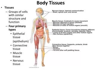
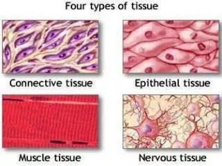
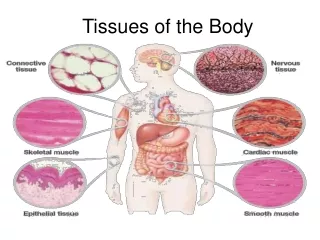
Body Tissues • Tissues • Groups of cells with similar structure and function • Four primary types • Epithelial tissue (epithelium) • Connective tissue • Muscle tissue • Nervous tissue
4-1 Four Types of Tissue • Epithelial Tissue • Covers exposed surfaces • Lines internal passageways • Forms glands • Connective Tissue • Fills internal spaces • Supports other tissues • Transports materials • Stores energy
4-1 Four Types of Tissue • Muscle Tissue • Specialized for contraction • Skeletal muscle, heart muscle, and walls of hollow organs • Neural Tissue • Carries electrical signals from one part of the body to another
Epithelial Tissues • Locations • Body coverings • Body linings • Glandular tissue • Functions • Protection • Absorption • Filtration • Secretion • Sensation
Epithelium Characteristics • Cells fit closely together and often form sheets (cell junctions) • The apical surface is the free surface of the tissue • The lower surface of the epithelium rests on a basement membrane (basal lamina) • Avascular (no blood supply) • Regenerate easily if well nourished
Tight junctions So close that are sometimes impermeable Adherens junctions Transmembrane linker proteins Desmosomes Anchoring junctions Filaments anchor to the opposite side Gap junctions Allow small molecules to move between cells Cell Junctions
Figure 4-2a Cell Junctions Tight junction Adhesion belt Terminal web Spotdesmosome Gapjunctions Hemidesmosome This is a diagrammatic view of an epithelial cell,showing the major types of intercellularconnections.
Figure 4-2b Cell Junctions Interlockingjunctionalproteins Tight junction Terminal web Adhesion belt A tight junction is formed by the fusion of the outer layers of two plasma membranes. Tight junctions prevent the diffusion of fluids and solutes betweenthe cells. A continuous adhesion belt lies deep to the tight junction. This belt is tied to the microfilaments of the terminal web.
Figure 4-2c Cell Junctions Embedded proteins(connexons) Gap junctions permit the free diffusion of ions and small molecules between two cells.
Figure 4-2d Cell Junctions Intermediatefilaments Cell adhesionmolecules (CAMs) Dense area Proteoglycans A spot desmosome tiesadjacent cells together.
Figure 4-2e Cell Junctions Clearlayer Basementmembrane Denselayer Hemidesmosomes attach a cell to extracellular structures, such as the protein fibers in the basement membrane.
Classification of Epithelia • Number of cell layers • Simple—one layer • Stratified—more than one layer Figure 3.17a
Classification of Epithelia • Shape of cells • Squamous • flattened • Cuboidal • cube-shaped • Columnar • column-like Figure 3.17b
Simple squamous Single layer of flat cells Functions in Absorption and diffusion Usually forms membranes Lines body cavities (Mesothelium) Lines hearts and capillaries (Endothelium) Simple Epithelia Lining of Artery
Figure 4-3a Squamous Epithelia Simple Squamous Epithelium LOCATIONS: Mesothelia lining ventral body cavities; endothelia lining heartand blood vessels; portions of kidney tubules (thin sections of nephron loops); inner lining of cornea; alveoli of lungs FUNCTIONS: Reduces friction; controls vessel permeability; performsabsorption and secretion Cytoplasm Nucleus Connective tissue LM 238 Lining of peritoneal cavity
Simple cuboidal Single layer of cube-like cells Common in glands and their ducts Forms walls of kidney tubules Covers the ovaries Functions in secretion and absorption Simple Epithelia
Figure 4-4a Cuboidal and Transitional Epithelia Simple Cuboidal Epithelium LOCATIONS: Glands; ducts;portions of kidney tubules; thyroidgland Connectivetissue FUNCTIONS: Limited protection,secretion, absorption Nucleus Cuboidalcells Basementmembrane Kidney tubule LM 650
Simple columnar Single layer of tall cells Often includes mucus-producing goblet cells Lines digestive tract Functions in absorption and secretion Simple Epithelia
Figure 4-5a Columnar Epithelia Simple Columnar Epithelium LOCATIONS: Lining ofstomach, intestine, gallbladder,uterine tubes, and collectingducts of kidneys Microvilli Cytoplasm FUNCTIONS: Protection,secretion, absorption Nucleus Basementmembrane Looseconnective tissue LM 350 Intestinal lining
Pseudostratified columnar Single layer, but some cells are shorter than others Often looks like a double layer of cells Sometimes ciliated, such as in the respiratory tract May function in absorption or secretion Cilia movement Simple Epithelia
Figure 4-5b Columnar Epithelia Pseudostratified Ciliated Columnar Epithelium LOCATIONS: Lining ofnasal cavity, trachea, andbronchi; portions of malereproductive tract Cilia Cytoplasm FUNCTIONS: Protection,secretion, move mucuswith cilia Nuclei Basementmembrane Looseconnective tissue Trachea LM 350
Stratified squamous Cells at the apical surface are flattened Found as a protective covering where friction is common Locations Skin Mouth Esophagus Stratified Epithelia • Protects against attacks • Keratin protein adds strength and water resistance
Figure 4-3b Squamous Epithelia Stratified Squamous Epithelium LOCATIONS: Surface of skin; lining of mouth, throat, esophagus, rectum, anus, and vagina FUNCTIONS: Provides physical protection against abrasion, pathogens, and chemical attack Squamoussuperficial cells Stem cells Basementmembrane Connectivetissue Surface of tongue LM 310
Stratified Epithelia • Stratified cuboidal—two layers of cuboidal cells - Found in sweat ducts and mammary glands • Rare in human body • Found mainly in ducts of large glands LOCATIONS: Lining of some ducts(rare) FUNCTIONS: Protection, secretion,absorption Lumenof duct Stratifiedcuboidalcells Basementmembrane Nuclei Connectivetissue
Stratified Epithelia • Stratified columnar—surface cells are columnar, cells underneath vary in size and shape • Rare in human body • Found mainly in ducts of large glands Stratified Columnar Epithelium LOCATIONS: Small areas ofthe pharynx, epiglottis, anus,mammary glands, salivarygland ducts, and urethra Looseconnective tissue Deeper basalcells FUNCTION: Protection Superficialcolumnar cells Cytoplasm Nuclei Basementmembrane
Transitional epithelium Shape of cells depends upon the amount of stretching Lines organs of the urinary system Stratified Epithelia • Tolerates repeated cycles of stretching and recoiling and returns to its previous shape without damage
Figure 4-4c Cuboidal and Transitional Epithelia Transitional Epithelium LOCATIONS: Urinarybladder; renal pelvis;ureters FUNCTIONS: Permitsexpansion and recoilafter stretching Epithelium(relaxed) Basement membrane Connective tissue andsmooth muscle layers LM 400 Empty bladder Epithelium(stretched) Basement membrane LM 400 Connective tissue andsmooth muscle layers LM 400 Full bladder Urinary bladder
Pap Smear (Papanicolaou Test) • Involves examining cells from the stratified squamous epithelium
Gland One or more cells responsible for secreting a particular product Two major gland types Endocrine gland Ductless since secretions diffuse into blood vessels (interstitial fluid) All secretions are hormones RELEASE hormones Exocrine gland Secretions empty through ducts to the epithelial surface Include sweat and oil glands PRODUCE hormones Glandular Epithelium
Classification of glands • By where they release their product • Exocrine: external secretion onto body surfaces (skin) or into body cavities • Endocrine: secrete messenger molecules (hormones) which are carried by blood to target organs; “ductless” glands • By whether they are unicellular or multicellular
Exocrine glandsunicellular or multicellular Unicellular: goblet cell scattered within epithelial lining of intestines and respiratory tubes Product: mucin mucus is mucin & water
Multicellular exocrine glands • Gland Structure • Multicellular glands • Structure of the duct • Simple (undivided) • Compound (divided) • Shape of secretory portion of the gland • Tubular (tube shaped) • Alveolar or acinar(blind pockets) • Relationship between ducts and glandular areas • Branched (several secretory areas sharing one duct)
Figure 4-7 A Structural Classification of Exocrine Glands Duct SIMPLE GLANDS Glandcells SIMPLETUBULAR SIMPLE COILEDTUBULAR SIMPLE BRANCHEDTUBULAR Examples: Examples: Examples: • Gastric glands • Merocrine sweat • Intestinal glands • Mucous glands glands of esophagus,tongue, duodenum SIMPLE ALVEOLAR(ACINAR) SIMPLE BRANCHEDALVEOLAR Examples: Examples: • Not found in adult; a • Sebaceous (oil) stage in developmentof simple branchedglands glands
Figure 4-7 A Structural Classification of Exocrine Glands COMPOUND GLANDS COMPOUND TUBULOALVEOLAR COMPOUNDTUBULAR COMPOUND ALVEOLAR(ACINAR) Examples: Examples: Examples: • Mucous glands (in mouth) • Salivary glands • Mammary glands • Bulbo-urethral glands (in • Glands of respiratory male reproductive system) passages • Testes (seminiferous • Pancreas tubules)
Examples of exocrine gland products • Many types of mucus secreting glands • Sweat glands of skin • Oil glands of skin • Salivary glands of mouth • Liver (bile) • Pancreas (digestive enzymes) • Mammary glands (milk)
Endocrine glands • Ductless glands • Release hormones into extracellular space • Hormones are messenger molecules • Hormones enter blood and travel to specific target organs
Connective Tissue • Found everywhere in the body • Includes the most abundant and widely distributed tissues • Functions - Structural support - Fluid transport - protection of organs - binds tissues together - energy storage - defense against microbes
Connective Tissue Characteristics • Specialized cells • Variations in blood supply • Some tissue types are well vascularized • Some have a poor blood supply or are avascular • Extracellular matrix • Non-living material that surrounds living cells\ • Makes up majority of tissue volume • Gives cells speciality