Download
1 / 16
160 likes | 169 Views
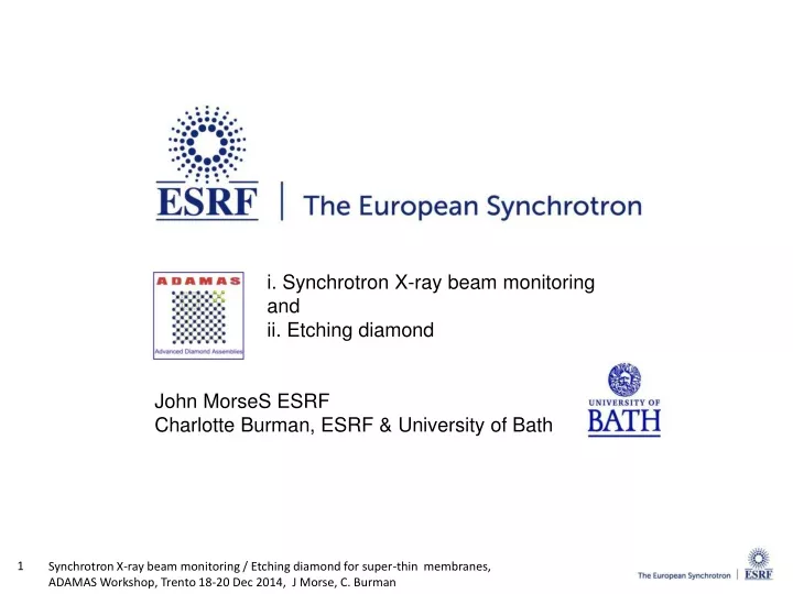
i. Synchrotron X-ray beam monitoring and ii. Etching diamond John MorseS ESRF Charlotte Burman, ESRF & University of Bath. Outline. 1. Synchrotrons and X-ray beam monitoring needs 2. diamond quadrant devices 3. CVD bulk and surface defects

E N D
i. Synchrotron X-ray beam monitoring and ii. Etching diamond John MorseS ESRF Charlotte Burman, ESRF & University of Bath
Outline 1. Synchrotrons and X-ray beam monitoring needs 2. diamond quadrant devices 3. CVD bulk and surface defects 4. diamond etching
The European Synchrotron Radiation Facility Third generation light source Location: Grenoble, France Cooperation: 20 countries Annual budget: ~100M€ Staff: 600 6.04Gev electron storage ring 844m circumference 32 straight sections 42 beamlines operating simultaneously some with 2 or 3 experimental stations X-ray beam energies ~1keV ...1MeV 10 Hz Booster Synchrotron 200 MeV Electron Linac User Availability: >98% of 250days/year Mean Time Between Failures: ~80 hours ~6000 annual user visits of duration ~few days ~2000 journal publications/year 3
diamond X-ray beam monitors: quadrant devices photo-ionization current readout → simple, compact devices • high purity diamond plate ~5…100µm thick, size ~10mm2 • low-Z metal 'blocking' contacts 20 ~ 100nm thick • externally applied bias field 0.5 ~ 5 Vµm-1 → full charge collection beam • absorption of small fraction of incident X-ray beam, diamond acts as solid state ‘ionization chamber’ • photo-electron thermalization range a few µm for <20keV X-rays DIAMOND • charge cloud drifts for ~ nanosecond in applied E field • transverse lateral thermal diffusion ~10µm during drift • beam 'center of gravity' determined by signal interpolation • -- difference/sum algorithm surface contact • signal currents can be measured with 'pulse averaging ' electrometers, or by narrow bandwidth • synchronized RF techniques • different signal measurement methods give different position response functions
Quadrant device with Keithley 485 electrometers (100msec integration), monochromatic beam ESRF ID09 beam on quadrant B 1000 550V current quad 2 (modulus nA) 100 beam on other quadrants (signal from beam halo?) 217V 10 138V 1 beam off ceramic package leakage e6 ELSC sample S361-1 0.1 17pA at +350V (390um thick, , 50µm quadrant isolation gap, TiW electrodes) -500 -400 -300 -200 -100 0 100 200 300 400 500 bias (volts) scan at 4V/sec signal variation with readout method SAME device measured at DESY-DORIS F1 (white bending magnet, Al filtered beam) with Libera RF readout system electrode ground bounce crosstalk Libera RF readout measures signal power in bandwidth ~5MHz at 500MHz synchrotron radiofrequency →only ‘fast' e, h charge drift induction signal (Ramo) within RF passband is measured → signal increases with bias as e, h carriers have not reached saturation drift velocity ( E fields ≤ 1.4Vµm-1)
1.E+00 Platinum electrodes M edge features: 1.E - 01 1.E - 02 1.E - 03 1.E - 04 1.E - 05 Gas ion chamber calibration 1.E - 06 Calorimetric calibration 1.E - 07 Fit, w = 13.4 +/ - 0.2 eV 1.E - 08 1.E - 07 1.E - 05 1.E - 03 1.E - 01 1.E+01 power Absorbed by Diamond (W) scCVD diamond responsivity with X-ray energy; linearity vs. X-ray flux data from e6 ELSC material responsivity fit diamond signal (Amps) J Morse et al, J. Synch. Rad 16 (2007) J. Bohon et al, J. Synch. Rad 17, (2010) J. Keister and J. Smedley, NIM A 606, (2009), 7 → linear current response demonstrated over 10 orders of magnitude !
threading dislocations → crystal strain visible with X-ray diffraction topography M.P. Gaukroger et al., Diam Relat. Mat. 2008 CVD bulk, surface defects or by polarized optical light transmission (birefringence) Surface damage from thinning/polishing laser cut high purity CVD overgrowth overgrowth with threading dislocations HPHT grown substrate crystal
Deep etching of diamond quadrant position monitors use signal interpolation, requires s/n ~103… 104 → need high uniformity of response across device active area ~10mm2 beam position and intensity monitoring measurement 'bandwidth' required is from zero …~1kHz → drift from polarization effects, and/or signal 'lag' cannot be tolerated (use of bias reversal very undesirable in this application) diamond polished plate ~50µm central area ArO etched to ~3µm metal electrodes ~50nm → need to remove polish-damaged sub-surface layer (several microns depth) plasma and ion beam etching techniques : ` planar removal of diamond surface with ~nanometer residual damage offers local area, masked etching to create robust, 'superthinned' (few µm) devices ~3um thick device tested at Soleil Synchrotron K Desjardins et al, J. Synchrotron Rad. (2014) 21 practical challenges: - etching processes are not inherently planarizing -need to avoid local etch pit formation at pre-existing bulk or surface defects -surface roughening related to existing polish damage of surface … and need process with ≥microns/hour etch rate
Deep etching - Project aims • To obtain adequate X-ray transparency for low energy X-ray beams (2~5 keV), diamonds must be ‘super-thinned’ to 5~20 µm. • High risk of plate edge chipping and breakage when processing to <50µm using scaife ‘abrasive’ polishing method. • Masked plasma etching can give robust • ‘window-framed’ membrane devices. See M.Pomorski, Appl. Phys. Lett. 103, 112106 (2013 Ion Beam Milling Inc. Argon etched • Consider/test different masking methods to delimit membrane area.
Masking techniques Laser machined polycrystalline diamond masks for plasma etching 4.5mm Vitreous carbon diamond holder
Deep etching - Project aims • To obtain adequate X-ray transparency for low energy X-ray beams (2~5 keV), diamonds must be ‘super-thinned’ to 5~20 µm. • High risk of plate edge chipping and breakage when processing to <50µm using scaife ‘abrasive’ polishing method. • Masked plasma etching can give robust • ‘window-framed’ membrane devices. See M.Pomorski, Appl. Phys. Lett. 103, 112106 (2013 Ion Beam Milling Inc. Argon etched • Consider/test different masking methods to delimit membrane area. • Compare different etchant gases and machine set-ups. • Determine how initial surface polish affects etch rates and final surface.
Plasma Diamond sample Plasma Etching techniques Electron cyclotron resonance plasma etching machine – Centre de Recherche Plasmas-Matériaux-Nanostructures, Grenoble, with Alexandre Bes. Inductively coupled plasma etching machine - PTA-Minatech, Grenoble, with Thierry Chevolleau and Thomas Charvolin.
Pure Oxygen etch result • Electron cyclotron resonance plasma etching Etch time: 120 minutes. Oxygen flow: 40sccm Pressure: 4.0mT Coil power: 2 x 600W Platen power: 150W Bias: ~ -142V
Argon/Oxygen etch result • Electron cyclotron resonance plasma etching Courtesy of Etienne Bustarret, Insitut Néel, CNRS, Grenoble Etch time: 60 minutes. Argon flow: 24sccm Oxygen flow: 4sccm Pressure: 7.0mT Coil power: 2 x 600W Platen power: 120W Bias: ~ -140V
Argon/Chlorine Etch Results Inductively coupled plasma etching machine - PTA-Minatech, Grenoble, with Thierry Chevolleau and Thomas Charvolin. Pre-etch surface RMS: 3.85nm Post-etch surface Etch time: 60 minutes, Argon flow: 25sccm, Chlorine flow: 40sccm. Lee, C.L et al. (2008) Diamond and Related Materials, 17 (7-10). pp. 1292-1296. RMS: 1.84nm
Conclusions Thank you. • Initial trials: surface quality (presence of damage pits on 'standard' e6 CVD samples) has major impact on final surface roughness and topology. • Pursuing trials with fine scaife polished HPHT 1b and CVD samples.