Download
1 / 50
500 likes | 506 Views
Digestion and alimentary tract in lower and higher organisms Main Parts of Human Digestive System Functions of the Liver Roles of Digestive enzymes during Digestion Modification of the alimentary tracts in herbivores. At the end of the period, students should be able to:

E N D
Digestion and alimentary tract in lower and higher organisms • Main Parts of Human Digestive System • Functions of the Liver • Roles of Digestive enzymes during Digestion • Modification of the alimentary tracts in herbivores
At the end of the period, students should be able to: • define alimentary canal & name its 5 main parts • define ingestion, absorption and egestion. • name the part of the alimentary canal where these occurs • identify all the main parts of the digestive system • list all the digestive enzymes in each part of the alimentary canal
Some invertebrates have a digestive cavity with a single opening • Sponges, the simplest invertebrates, obtain food by filtering microscopic organisms from the surrounding water. • Individual cells phagocytose the food particles, and digestion is intracellular within food vacuoles. • Wastes are egested into the water that continuously circulates through the sponge body.
Most invertebrates, and all vertebrates, have a tube-within-a-tube body plan. • The body wall forms the outer tube. • The inner tube is a digestive tract with two openings, sometimes referred to as a complete digestive system. • Food enters through the mouth, and undigested food is eliminated through the anus. • The mixing and propulsive movements of the digestive tract are referred to as motility. • The propulsive activity characteristic of most regions of the digestive tract is peristalsis, waves of muscular contraction that push the food in one direction. • More food can be taken in while previously eaten food is being digested & absorbed farther down the digestive tract. • In a digestive tract with two openings, various regions of the tube are adapted to perform specific function
Lower organisms such as Sponges, Amoeba, Paramecium have no special digestive tracts at all. • They feed on microscopic organisms by phagocytosis, then digestive the food particles intracellularly within the food vacuoles. • Excess water and waste products are then liberated into the surrounding water.
Cnidarians such as hydra, jellyfish feed on cylops, annelids, crustaceans, insect larvae. Their mouth serves for both ingestion and egestion as they lack anus. • Cnidarians use tentacles to capture and direct the prey. The prey is killed extracellularly by the action of digestive enzymes from gland cells of gastrodermis. • Intracellular digestion occurs in the gastrovascular cavity and excess materials are egested via the anus.
Ingestion: placing food into the mouth (entry of food in the digestive system), • Digestion: This can be defined as the mechanical, physical and chemical breakdown, mastication and the mixing of food into into simple form that is easily available for absorption by the body.
Absorption: of nutrients from the digestive system to the circulatory and lymphatic capillaries through osmosis, active transport & diffusion. • Egestion: Removal of undigested materials from the digestive tract through defecation.
The liver, pancreas, and, in terrestrial vertebrates, the salivary glands are accessory glands that secrete digestive juices into the digestive tract.
From inside out, the layers of the wall are the mucosa, submucosa, muscle layer, and visceral peritoneum. • Although various regions differ somewhat in structure, the layers are basically similar throughout the digestive tract . • The mucosa, a layer of epithelial tissue and underlying connective tissue, lines the lumen (inner space) of the digestive tract. • In the stomach and intestine, the mucosa is greatly folded to increase the secreting & absorbing surface. • Surrounding the mucosa is the submucosa, a connective tissue layer rich in blood vessels, lymphatic vessels, and nerves.
The liver is the heaviest organ in the body and is one of the largest. • Lies in the upper right part of your belly under the ribs and is responsible for functions vital to life. • The main function is to process nutrients from food, make bile, remove toxins from the body and build proteins. • It's easy to see how inflammation of the liver, or hepatitis, interferes with these important functions and can lead to poor health. • Fortunately, the liver is extremely resilient and most cases of liver inflammation don't even come to medical attention, but in cases of severe liver disease, there can be serious interruption of these essential liver functions.
FUNCTIONS OF THE LIVER • Deamination is the removal of an amine group from a molecule. Enzymes that catalyse this reaction are called deaminases. • In the human body, deamination takes place primarily in the liver, however glutamate is also deaminated in the kidneys. • Deamination is the process by which amino acids are broken down if there is an excess of protein intake. The amino group is removed from the amino acid and converted to ammonia. • The rest of the amino acid is made up of mostly carbon and hydrogen, and is recycled or oxidized for energy. Ammonia is toxic to the human system, and enzymes convert it to urea or uric acid by addition of carbon dioxide molecules (which is not considered a deamination process) in the urea cycle, which also takes place in the liver. Urea and uric acid can safely diffuse into the blood and then be excreted in urine.
The liver is considered a gland—an organ that secretes chemicals—because it produces bile, a substance needed to digest fats. • Bile’s salts break up fat into smaller pieces so it can be absorbed more easily in the small intestine. • In addition to producing bile, the liver: • Detoxifies the blood to rid it of harmful substances such as alcohol and drugs • Stores some vitamins and iron • Stores the simple sugar glucose • Converts stored sugar to usable sugar when the body’s sugar (glucose) levels fall below normal. • Breaks down hemoglobin as well as insulin and other hormones • Converts ammonia to urea, which is vital in metabolism • Destroys old red blood cells
Bile is a thick, green-yellow fluid that the liver produces to help digest food, especially fat, as it passes from the stomach to the intestines. • Bile is stored in a nearby sac called the gall bladder after its production. • When a person eats a meal heavy in fat, the body will use its store of bile to help break down the fats for digestion.
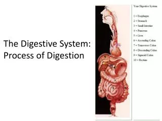
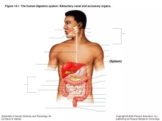
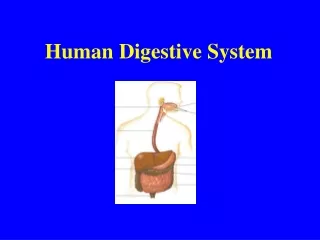
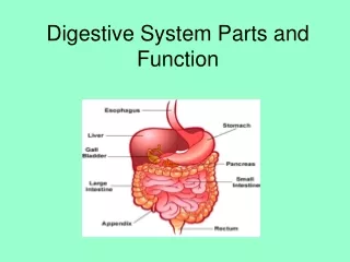
5 MAIN PARTS OF DIGESTIVE SYSTEM • MOUTH • EOSOPHAGUS • STOMACH • SMALL INTESTINE • LARGE INTESTINE
THE MOUTH • Here lies the tongue between the dental arches and the cheek. • Produces alkaline saliva from salivary glands • Secretes Ptyalin which convert starch to maltose (chemical digestion) • Teeth cut masticate the food into smaller pieces • Mouth opens into muscular pharynx • During swallowing, the epiglottis flaps down to cover the trachea to prevent choking.
OESOPHAGUS • It is a muscular band of muscles capable of contracting and relaxing • Food moves by peristalsis • Peristalsis occurs throughout the walls of the alimentary gut which are muscular • No enzyme is secreted here
OESOPHAGUS • The oesophagus is a muscular tube through which food is carried from the pharynx to the stomach. • The oesophagus also has to accommodate a wide variety of food and drink (hot, cold, spicy etc). • The oesophagus has a stratified squamous epithelial lining(SE) which protects the oesophagus from trauma.
Food is mechanically and enzymatically digested in the stomach • The entrance to the large, muscular stomach is normally closed by a ring of muscle at the lower end of the esophagus. • When a peristaltic wave passes down the esophagus, the muscle relaxes and the bolus enters the stomach • When empty, the stomach is collapsed and shaped almost like a hot dog. • Folds of the stomach wall, called rugae, give the inner lining a wrinkled appearance. • As food enters, the rugae gradually smooth out, which expands the capacity of the stomach to more than a liter. • The stomach is lined with a simple, columnar epithelium that secretes large amounts of mucus.
Tiny pits mark the entrances to the millions of gastric glands, which extend deep into the stomach wall. • Parietal cells in the gastric glands secrete hydrochloric acid and intrinsic factor, a substance needed for adequate absorption of vitamin B12. • Chief cells in the gastric glands secrete pepsinogen, an inactive enzyme precursor. • When it comes in contact with the acidic gastric juice in the stomach, pepsinogen is converted to pepsin, the main digestive enzyme of the stomach. • Pepsin hydrolyzes proteins, converting them to short polypeptides • Several protective mechanisms prevent the gastric juice from digesting the wall of the stomach. • Cells of the gastric mucosa secrete an alkaline mucus that coats the stomach wall and neutralizes the acidity of the gastric juice along the lining. • In addition, the epithelial cells of the lining fit tightly together, preventing gastric juice from leaking between them and into the tissue beneath
Food enters the stomach from the esophagus when the muscles of the cardiac sphincter on the upper region of the stomach are relaxed. • Contraction of the muscular walls cause churning of food • Secrete gastric juice (work best in acidic medium) which contain 2 enzymes. About two litres of gastric juices is secreted daily by the gastric glands • Food enters the stomach from the esophagus when the muscles of the cardiac sphincter on the upper region of the stomach are relaxed.
The chief cells of gastric glands produces two types of enzymes namely: pepsin and rennin. • Pepsinogen is initially secreted but later activated to form pepsin by the action of dilute hydrochloric acid. • Pepsin converts protein to peptones • Rennin converts soluble milk, caseinogen to casein especially in infants • Apart from activating pepsinogen to form pepsin, dilute HCl in the stomach helps maintain the pH of the gastric juice, also has antiseptic property capable of killing pathogens that may be present in the food. • Food exits the stomach when the muscles of the pyloric sphincter are relaxed
Sometimes, these protective mechanisms malfunction and part of the stomach lining is digested, leaving an open sore, or peptic ulcer. • Such ulcers often occur in the duodenum and sometimes in the lower part of the esophagus. • The bacterium Helicobacter pylori has been implicated as a causative factor in ulcers • Helicobacter pylori infects the mucus-secreting cells of the stomach lining, decreasing the protective mucus, which can lead to peptic ulcers or cancer. • This type of infection responds to antibiotic therapy.
The stomach is a 'J'-shaped organ, with two openings- the oesophageal and the duodenal. • Has four regions- the cardia, fundus, body and pylorus. • Each region performs different functions; the fundus collects digestive gases, the body secretes pepsinogen and hydrochloric acid, & the pylorus is responsible for mucous, gastrin and pepsinogen secretion. • Stomach has 5 major functions; • Temporary food storage of food • Control the rate at which food enters the duodenum • Acid secretion and antibacterial action • Fluidisation of stomach contents • Preliminary digestion with pepsin, lipases etc.
Divided into 3 parts viz Duodenum, Jejunum & Ileum • Food enters duodenum(the first part) • Pancreas secretes alkaline pancreatic juice which contains 3 enzymes viz • Amylopsin (starch to maltose) • Trypsin (protein to polypeptides) • Lipase (fats to fatty acids & glycerol)
Presence of numerous typically finger-like processes called Villi that project from the surface of the mucosa into the lumen. • The villi are greatest and most numerous for a given area of the duodenum. • Each villus consist of a central lymphatic vessel called Lacteal which may occasionally be doubled. • The villi may sometimes be leaf-like or flattened. • Presence of numerous depressions called Crypts that project from the surface of the mucosa into the lumen.
REGULATION OF DIGESTIVE SECRETIONS BY HORMONES • It is important to note that hormones such as gastrin, secretin, cholecystokinin & enterogastrone a.k.a gastric inhibitory peptide (GIP) play vital roles in digestion. • Gastrin is secreted in the mucosa wall of the stomach • Secretin, cholecystokinin and GIP are all secreted in the mucosa walls of the duodenum.
REGULATION OF DIGESTIVE SECRETIONS BY HORMONES • Gastrin signals gastric glands to release pepsinogen • Secretin stimulates the release of sodium bicarbonate by the pancreas & bile to neutralize the acidic medium of chyme in the stomach • Cholecystokinin stimulates the contraction of the gall bladder to release bile into the small intestine & the release of pancreatic enzymes. • Enterogastrone inhibits peristalsis in the stomach in order to slow down the entry of chyme into the duodenum.
Most enzymatic digestion takes place in the small intestine • Digestion of food is completed in the small intestine, and nutrients • are absorbed through its wall. • The small intestine, which is 5 to 6 m (about 17 ft) in length, has three regions: the duodenum, jejunum, and ileum. • Most chemical digestion takes place in the duodenum, the first portion of the small intestine, not in the stomach. • Bile from the liver and enzymes from the pancreas are released into the duodenum and act on the chyme. • Then enzymes produced by the epithelial cells lining the duodenum catalyze the final steps in the digestion of the major types of nutrients.
The lining of the small intestine appears velvety because of its millions of tiny fingerlike projections, the intestinal villi (sing., villus). • The villi increase the surface area of the small intestine for digestion and absorption of nutrients. • The intestinal surface is further expanded by microvilli, projections of the plasma membrane of the simple columnar epithelial cells of the villi. • About 600 microvilli protrude from the exposed surface of each cell, giving the epithelial lining a fuzzy appearance when viewed with the electron microscope. • If the intestinal lining were smooth, like the inside of a water pipe, food would zip right through the intestine & many valuable nutrients would not be absorbed. • Folds in the wall of the intestine, the villi, & microvilli together increase the surface area of the small intestine by about 600 times. • The lining of the small intestine of an adult human, if spread out, its surface would approximate the size of a tennis court.
Liver produces watery bile which emulsify fats into tiny droplet • Bile contains no enzymes • Help add water to chyme • Chyme turns to chyle as it passes
There are also intestinal juice which contains • Maltase (Maltose to glucose + glucose) • Lactase (Lactose to glucose + galactose) • Sucrase (Sucrose to glucose+ fructose) • Erepsin : Polypeptides to amino acids • Lipase : Fats to carboxylic acids + glycerol
Polysaccharides, such as starch and glycogen, are important components of the food ingested by most animals. • The glucose units of these large molecules are connected by glycosidic bonds . • These bonds are hydrolyzed by amylases that digest polysaccharides to the disaccharide maltose. • Although amylase can split the a-glycosidic linkages present in starch and glycogen, it cannot split the b-glycosidic linkages present in cellulose. • Amylase cannot split the bond between the two glucose units of maltose. • Enzymes produced by the cells lining the small intestine break down disaccharides such as maltose to monosaccharides. • Hydrolysis occurs while the disaccharides are being absorbed through the epithelium of the small intestine .
Pepsin, trypsin, and chymotrypsin break certain internal peptide bonds of proteins and polypeptides. • Carboxypeptidase removes amino acids with free terminal carboxyl groups from the end of polypeptide chains. • Dipeptidases released by the duodenum then split the small peptides to amino acids. • Consequently, the products of lipid digestion include monoacylglycerols (monoglycerides) and diacylglycerols (diglycerides) as well as glycerol and fatty acids. • Undigested triacylglycerols also remain, some of which are absorbed without digestion.
Facilitated by villi, a finger-like projection& blind lymphatic tube called Lacteal • Villi is surrounded by network of blood capillaries (vascularised) • Lymphatic vessels eventually empty their content into the blood vessels • Protein is digested to amino acids, CHO to glucose and lipids to fatty acids and glycerol. • Amino acids & glucose are transported to the liver by the hepatic portal vein
Villi increase the surface area of small intestine: absorption of food is facilitated
Undigested food passes into colon • More water absorbed • This makes it more concentrated • Faeces are stored in rectum • Leaves the body through the anus
The Large Intestine • This is divided into six regions namely caecum, colon, rectum and anus. • Colon is further subdivided into ascending, transverse & descending colons. • Rectum is the last portion of the large intestine& terminates in the anal sphincter. • A structure called the vermiform appendix extends from blind end of rectum & its inflammation causes appendicitis. • Colon plays fewer roles in digestion but it is important for reabsorption of fluids which benefit the body. • The large intestine consist mainly of columnar cells specialized for water & sodium reabsorption and mucus-secreting goblet cells to aid the passage of faeces.
Ruminants have the most elaborate adaptations for herbivorous diets & have a 4-chambered stomach: Rumen, Reticulum, Omasum & Abomasum. • 3 of the 4 ruminant stomach compartments (rumen, reticulum & omasum) make up the forestomach & are extension of the lower oesophagus. • The rumen, the first of the forestomach chambers, stores and processes plant material. • Huge numbers of these bacteria are present in the rumen and the reticulum: with each gram of rumen fluid contains 10 - 50 billion bacteria
RUMINANTS DIGESTIVE TRACTS • Ruminant first chew & swallows its food in the mouth, it enters the rumen & reticulum where symbiotic bacteria act on the cellulose-rich meal. • Ruminants tend to swallow their food quickly, with only minimal mastication. • When ruminants is resting after grazing, it regurgitates this partially chewed food, rechews it, then swallows it again making it more accessible to further bacteria actions. • This process is called “chewing the cud” or rumination. • Depending on the amount of fibre in their food, cow may spend 3-6 hours per day chewing their cud.
As by-products of their metabolism, the bacteria secrete fatty acids. • The cow periodically regurgitates & rechews the cud(4) which further breaks down the fibres • The cow then reswallows the cud which moves to the omasum, where the water is removed. • The cud finally passes to the abomasum for digestion, • The diet from which a ruminant actually absorb its nutrients is more richer than the hay or grass the animal originally eats. • Infact, ruminants obtain many of its nutrients by digesting the symbolic bacteria which reproduce rapidly enough in the rumen..
Thank you for listening Dr. O. A. Iyiola