Download
1 / 20
380 likes | 3.31k Views
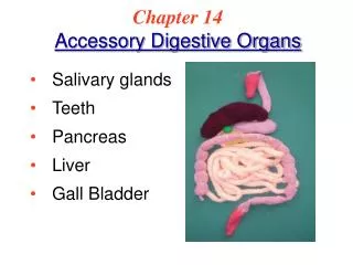
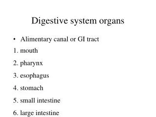
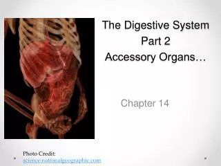
Figure 14.1 The human digestive system: Alimentary canal and accessory organs. (Spleen). Figure 14.1 The human digestive system: Alimentary canal and accessory organs. Parotid gland. Mouth (oral cavity). Sublingual gland. Salivary glands. Submandibular gland. Tongue. Pharynx.

E N D
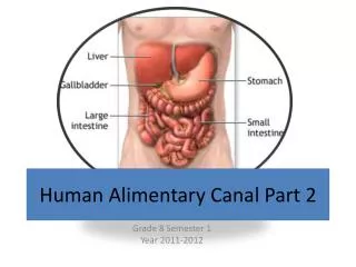
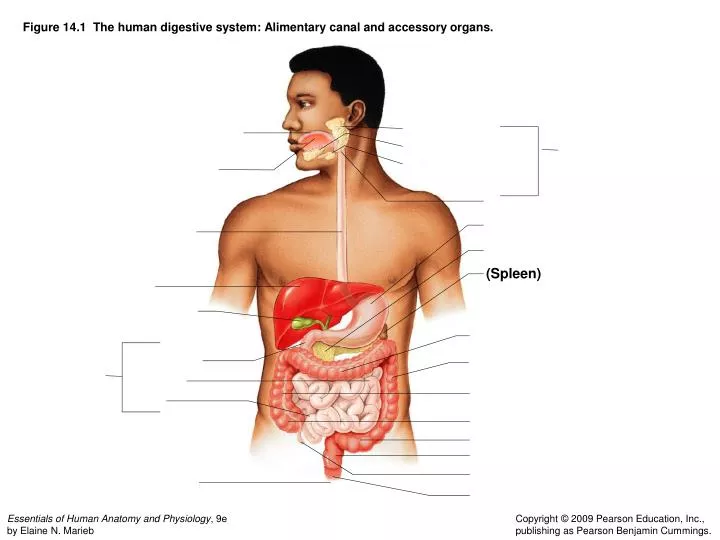
Figure 14.1 The human digestive system: Alimentary canal and accessory organs. (Spleen)
Figure 14.1 The human digestive system: Alimentary canal and accessory organs. Parotid gland Mouth (oral cavity) Sublingual gland Salivary glands Submandibular gland Tongue Pharynx Stomach Esophagus Pancreas (Spleen) Liver Gallbladder Large intestine • Transverse colon Small intestine • Duodenum • Descending colon • Jejunum • Ascending colon • Ileum • Cecum • Sigmoid colon • Rectum • Appendix Anus • Anal canal
Figure 14.2a Anatomy of the mouth (oral cavity). (a) Sagittal view of the oral cavity and pharynx. Trachea (a)
Figure 14.2a Anatomy of the mouth (oral cavity). (a) Sagittal view of the oral cavity and pharynx. Nasopharynx Hard palate Soft palate Oral cavity Uvula Lips (labia) Palatine tonsil Vestibule Lingual tonsil Oropharynx Lingual frenulum Epiglottis Tongue Laryngopharynx Hyoid bone Esophagus Trachea (a)
Figure 14.2b Anatomy of the mouth (oral cavity). (b) Anterior view of the oral cavity. (b)
Figure 14.2b Anatomy of the mouth (oral cavity). (b) Anterior view of the oral cavity. Upper lip Gingivae (gums) Hard palate Soft palate Uvula Palatine tonsil Oropharynx Tongue (b)
Figure 14.3 Basic structure of the alimentary canal wall. (visceral peritoneum) Nerve Gland in mucosa Artery Duct of gland outside alimentary canal Vein
Figure 14.3 Basic structure of the alimentary canal wall. Visceral peritoneum Submucosal glands Mucosa Surface epithelium Lamina propria Muscle layer Submucosa Muscularis externa Longitudinal muscle layer Circular muscle layer Serosa (visceral peritoneum) Nerve Gland in mucosa Lumen Artery Mesentery Duct of gland outside alimentary canal Vein Lymph nodule
Figure 14.4a Anatomy of the stomach. (a) Diagram of the gross internal anatomy (frontal section). (a)
Figure 14.4a Anatomy of the stomach. (a) Diagram of the gross internal anatomy (frontal section). Cardioesophageal sphincter Fundus Esophagus Muscularis externa Serosa Longitudinal layer Circular layer Body Oblique layer Lesser curvature Rugae of mucosa Pylorus Greater curvature Duodenum Pyloric sphincter (valve) Pyloric antrum (a)
Figure 14.4c Anatomy of the stomach. (c) Enlarged view of gastric pits and glands (longitudinal section). (c)
Figure 14.4c Anatomy of the stomach. (c) Enlarged view of gastric pits and glands (longitudinal section). Gastric pits Surface epithelium Pyloric sphincter Mucous neck cells Parietal cells Gastric glands Chief cells (c)
Figure 14.7a Structural modifications of the small intestine. (a) Small intestine. Blood vessels serving small intestine (a) Small intestine
Figure 14.7a Structural modifications of the small intestine. (a) Small intestine. Blood vessels serving small intestine Muscle layers Large circular folds (plicae circulares) Villi (a) Small intestine
Figure 14.7b Structural modifications of the small intestine. (b) Villi. Vein Artery Submucosa (b) Villi
Figure 14.7b Structural modifications of the small intestine. (b) Villi. One villus Lacteal Blood capillaries Vein Artery Submucosa (b) Villi
Figure 14.8 The large intestine. Left colic (splenic) flexure Transverse mesocolon Right colic (hepatic) flexure Transverse colon Descending colon Haustra Ascending colon IIeum (cut) Cut edge of mesentery IIeocecal valve Teniae coli Sigmoid colon Cecum Appendix Rectum Anal canal External anal sphincter
Figure 14.11 Schematic summary of gastrointestinal tract activities. Food Pharynx Esophagus • Chewing (mouth) • Churning (stomach) • Segmentation (small intestine) • Swallowing (oropharynx) Chemical digestion • Peristalsis (esophagus, stomach, small intestine, large intestine) Stomach Lymph vessel Small intestine Blood vessel Large intestine Mainly H2O Feces Anus
Figure 14.11 Schematic summary of gastrointestinal tract activities. Food Ingestion Mechanical digestion Pharynx Esophagus • Chewing (mouth) • Churning (stomach) Propulsion • Segmentation (small intestine) • Swallowing (oropharynx) Chemical digestion • Peristalsis (esophagus, stomach, small intestine, large intestine) Stomach Absorption Lymph vessel Small intestine Blood vessel Large intestine Mainly H2O Feces Anus Defecation